icon

Cystectomy: What You Need to Know

A cystectomy is a surgical procedure for the removal of the urinary bladder. In some cases, surrounding tissues and organs in the pelvis are also removed, depending on the underlying health condition being treated.

This surgery is most commonly performed to treat bladder cancer but may also be indicated for other severe bladder conditions, such as significant bladder dysfunction.

Types of Cystectomy

Cystectomy procedures vary mainly by the extent of bladder removal and the surgical method used. Here are the main types of cystectomy:

Partial Cystectomy

In a partial cystectomy, only a portion of the bladder is removed. This procedure is suitable for patients whose cancer is located in one area of the bladder and has not spread to the muscle layer.

Radical Cystectomy

Radical cystectomy involves the removal of the entire bladder along with nearby lymph nodes, part of the urethra, and, depending on the patient’s sex, potentially the prostate and seminal vesicles in men, or the uterus, ovaries, and part of the vagina in women.

Robotic Cystectomy

Robotic cystectomy is a minimally invasive approach to radical cystectomy, performed using robotic surgical systems. This method offers the surgeon enhanced precision, flexibility, and control.

Reasons for Cystectomy

Here are the common reasons for undergoing a cystectomy:

Bladder Cancer: Bladder cancer is the most common indication for a cystectomy.

Cystectomy may also be considered for certain severe non-cancerous conditions when other treatments have failed, including:

Interstitial cystitis: A chronic condition causing bladder pressure, bladder pain and sometimes pelvic pain.

Neurogenic bladder: Dysfunction of the urinary bladder due to disease of the central nervous system or peripheral nerves involved in the control of urination.

Bladder trauma: Severe injuries to the bladder that cannot be repaired by other surgical means.

The Cystectomy Procedure

The cystectomy procedure involves several steps, such as:

Preoperative Preparation

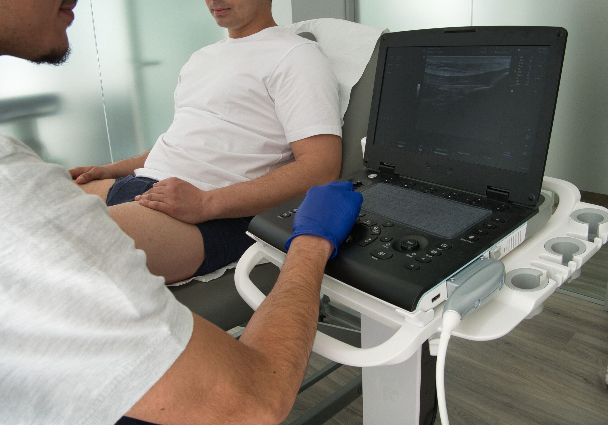
Patients undergoing cystectomy typically undergo a thorough preoperative assessment, including blood tests, imaging studies, and sometimes cardiac and pulmonary evaluations, to ensure they are fit for surgery.

Surgical Procedure

  1. Anaesthesia: Cystectomy is performed under general anaesthesia.
  2. Incision: For a traditional open cystectomy, a single large incision is made across the abdomen to access the bladder. For robotic and laparoscopic cystectomies, several small incisions are made to accommodate surgical instruments and a camera.
  3. Removal of the Bladder: The bladder, and often surrounding lymph nodes and other tissues, are removed. The extent of tissue removal depends on the underlying condition and the type of cystectomy.
  4. Urinary Diversion: After removing the bladder, surgeons must create a new way for urine to exit the body. Options include:
  • Ileal conduit urinary diversion: Using a piece of the intestine to create a conduit outside the abdomen.
  • Continent urinary reservoir: Creating a reservoir that can be emptied by inserting a catheter through the abdomen.

Postoperative Care

Postoperative care involves monitoring for signs of infection, managing pain, and ensuring proper surgical site healing. Patients may need to stay in the hospital for several days to a few weeks, depending on the surgery’s complexity and overall health.

Potential Risks and Complications

Cystectomy, like any major surgery, carries potential risks and complications, including:

Immediate Postoperative Risks

Bleeding: Significant blood loss can occur during or after the surgery, sometimes necessitating blood transfusions.

Infection: Surgical sites, urinary diversions, or the bloodstream can become infected.

Anaesthetic Complications: Reactions to anaesthesia can affect cardiac and respiratory functions.

Long-term Complications

Urinary Tract Infections (UTIs): Patients with any form of urinary diversion are at increased risk of UTIs.

Bowel Obstruction: Scar tissue from surgery can cause obstructions in the intestines.

Ureteral Stricture: Narrowing of the ureters, which can lead to kidney problems.

Surgical Failure or Recurrence of Disease

Cancer Recurrence: There is always a risk that cancer can recur, even after the removal of the bladder and surrounding tissues.

Complications from Urinary Diversion: Complications arising from urinary diversions, such as issues with the constructed urinary pathways, may require additional surgeries.

Conclusion

Cystectomy is a surgical procedure to manage and treat bladder cancer or other severe bladder conditions. The decision to undergo a cystectomy involves careful consideration of the benefits and risks associated with the procedure. Choosing the right surgical option and understanding the potential outcomes are important for managing one’s health effectively.

For more detailed information about cystectomy, or to discuss whether this surgical option might be appropriate for your condition, consider scheduling a consultation with our clinic. Our team is ready to provide you with comprehensive advice and support tailored to your health needs.

Recovery Timeline For Phimosis Surgery: A Urologist Surgeon’s Guide

Phimosis is the inability to retract the foreskin over the glans penis. It can cause discomfort, urinary difficulties, and hygiene issues. This condition can be present from birth or develop later in life, and while it’s common in infants and young children, it can also affect adults.

This procedure involves the removal of the foreskin, tailored to the severity of the condition and patient preferences.

Purpose of Surgery: The surgery aims to alleviate symptoms and prevent complications such as paraphimosis, where the foreskin becomes trapped behind the glans. The procedure typically lasts 30 to 60 minutes.

Surgical Goals: The main objectives are to reduce immediate discomfort, avoid future infections, and ensure normal urinary function.

Immediate Post-Surgical Care

Immediately following phimosis surgery, the primary focus is on managing pain and preventing infection.

Pain Management

Analgesics: Patients are usually prescribed pain relief medications such as paracetamol or ibuprofen. These help manage discomfort and should be taken as directed.

Ice Packs: Applying ice packs to the surgical area can help reduce swelling and alleviate pain.

Hygiene

Cleaning the Area: The surgical site should be kept clean and dry. Patients are advised to gently wash the area with warm water and avoid using any harsh soaps or chemicals.

Dressing Changes: If a dressing was applied, it might need to be changed regularly. The urologist will give specific instructions on how often and how to change the dressing safely.

Activity Restrictions

Rest: Significant physical activity should be avoided to prevent aggravation of the surgical site. Avoidance of Sexual Activity: Patients are advised to abstain from sexual activity until the site is fully healed, which is usually around 4 to 6 weeks post-surgery.

Monitoring for Complications

Signs of Infection: Any signs of infection, such as increased redness, swelling, pus, or fever, should be reported to a doctor immediately.

Unusual Pain or Bleeding: While some pain is normal, excessive pain or bleeding should be promptly evaluated.

First Week of Recovery

The first week after phimosis surgery is critical for healing and monitoring. Patients should manage discomfort, maintain hygiene, and observe any signs indicating complications.

Pain and Swelling

Continued Pain Management: Pain typically diminishes over the first week but may require ongoing management with prescribed analgesics.

Swelling: Some swelling is normal. Continuing with intermittent ice pack applications can help reduce this.

Observation

Observation of the Surgical Site: It’s important to check the site daily for signs of infection or other complications.

Physical Activity

Limited Mobility: Patients are advised to limit walking and avoid strenuous activities. Light walking around the house is encouraged to promote blood circulation.

Diet and Hydration

Hydration: Keeping hydrated helps the body heal and reduces the risk of urinary tract infections.

Nutritious Diet: Eating well-balanced meals rich in vitamins and proteins can aid in the healing process.

Subsequent Weeks of Recovery

As the patient moves beyond the initial week post-phimosis surgery, the focus shifts to continued healing, increased mobility, and a gradual return to normal activities.

Reduced Pain and Swelling

Decreasing Discomfort: Pain and swelling should gradually decrease.

Wound Healing

Improvement in Surgical Site: The surgical site should start showing signs of healing with reduced redness and swelling.

Suture Absorption or Removal: If non-absorbable sutures were used, they might be removed during a follow-up visit, typically 2-3 weeks post-surgery.

Increased Mobility

Resumption of Activities: Patients can resume more normal activities, such as returning to work and light exercise, depending on their comfort level and the nature of their job.

Gradual Return to Sexual Activity: If healing is progressing well and with the doctor’s approval, sexual activities can gradually resume, typically after 4-6 weeks, ensuring no discomfort or pain.

Continuous Hygiene and Care

Ongoing Cleaning: It remains important to keep the surgical area clean and monitor for any signs of infection.

Regular Check-Ups: Regular visits to a urologist may continue to ensure optimal healing and address any concerns.

Long-Term Care and Monitoring

Long-term care and monitoring after phimosis surgery are essential to ensure the health and functionality of the penis are maintained.

  • Regular Self-Examination: Patients should regularly examine the surgical area to ensure no new symptoms such as redness, swelling, or discharge have developed.
  • Sexual Function: It’s important to monitor any changes in sexual function, including sensitivity and satisfaction. Any concerns should be discussed with a urologist.
  • Follow-Up Visits: Depending on the person’s case, some patients may require annual visits to a urologist to ensure ongoing health and address any late complications.

Conclusion

Recovery from phimosis surgery varies among people but typically follows a clear timeline that begins with immediate post-surgical care and extends through several weeks of recovery and long-term monitoring.

The key to a successful recovery involves diligent adherence to postoperative care instructions, regular monitoring for complications, and maintaining good hygiene practices.

By understanding and following the outlined recovery phases, patients can expect a satisfactory outcome from phimosis surgery, with improved symptoms and enhanced quality of life.

Phimosis Treatment Options: A Guide For Prospective Patients

Phimosis is a medical condition where the foreskin cannot be fully retracted over the glans penis.

For some, phimosis presents minimal or no symptoms, while for others, it can lead to discomfort, difficulty in maintaining hygiene, pain during urination, and complications during sexual activity.

Understanding Phimosis

Phimosis is categorised into two types: physiological and pathological.

In contrast, pathological phimosis occurs due to scarring, infection, or inflammation and does not resolve on its own, often necessitating medical treatment.

Pathological phimosis requires active treatment to prevent further complications such as urinary infections, pain, and issues with sexual function.

Non-Surgical Treatment Options

Non-surgical approaches are often the first line of treatment for phimosis, particularly when the condition is not severe or caused by scarring.

Steroid Creams

Topical steroid creams are commonly prescribed to treat phimosis. These creams are applied directly to the foreskin.

Mechanism: The active compounds in steroid creams help reduce inflammation and promote the elasticity of the skin, making it easier to retract the foreskin over time.

Duration: Treatment usually lasts for a few weeks. During this period, regular application of the cream and engagement in gentle stretching exercises are advised to enhance treatment effectiveness.

Gentle Stretching

In conjunction with steroid creams, or as a standalone method, gentle manual stretching of the foreskin can also be an effective treatment.

Technique: This involves the patient or a caregiver gently pulling back the foreskin incrementally to increase its elasticity.

Precautions: It is important to be gentle and avoid forceful retraction, which can lead to tears, scars, and further complications.

Guidance: Performing these exercises under the guidance of a urologist ensures that they are done correctly and safely, minimising the risk of injury.

Surgical Treatment Options

When non-surgical treatments are ineffective or unsuitable, surgical interventions become necessary. These options offer a more definitive resolution to phimosis, especially in severe cases.

Circumcision

Circumcision is the most common surgical procedure for treating phimosis and involves the complete removal of the foreskin.

Procedure: The surgery is performed under local or general anaesthesia, depending on the patient’s age and overall health. The foreskin is surgically removed, exposing the glans permanently.

Recovery: Post-operative care includes keeping the area clean and dry, avoiding physical activities that might irritate or impact the surgical site, and managing pain with prescribed medications. Full recovery is typically expected within a few weeks.

Considerations: Circumcision is a permanent solution often recommended in cases where phimosis has led to recurrent infections or other complications.

Preputioplasty

Preputioplasty offers an alternative to circumcision by preserving the foreskin while relieving its tightness.

Procedure: This surgery involves making one or more longitudinal incisions on the foreskin, which are then stitched in a transverse fashion to widen the opening.

Recovery: The recovery period is generally shorter than that of circumcision, with most patients experiencing less discomfort as well.

Considerations: Preputioplasty is less invasive than circumcision and is suitable for patients who wish to retain their foreskin. It is particularly considered when cosmetic or cultural reasons are important.

Frenuloplasty

Frenuloplasty specifically addresses a short frenulum, which can contribute to phimosis.

Procedure: The procedure involves making an incision in the frenulum to lengthen it, which helps in reducing the tightness of the foreskin

Recovery: Recovery from frenuloplasty is typically quick, with most patients returning to normal activities quickly.

Considerations: This surgery is recommended when the frenulum is the primary cause of the tightness and is often performed alongside other treatments like steroid creams or preputioplasty.

Risks and Considerations of Treatment

Choosing the proper treatment for phimosis involves understanding each option’s potential risks and considerations. It’s important for patients to discuss these aspects with a urologist to make an informed decision.

Non-Surgical Risks

Risks and Considerations of Treatment

Skin Irritation or Allergic Reactions: Use of steroid creams, while generally safe, may lead to skin irritation or allergic reactions in some people.

Micro-tears: Overzealous stretching can lead to micro-tears in the foreskin, which may cause scarring and potentially worsen the condition.

Surgical Risks

Infection: Like all surgical procedures, phimosis surgery carries a risk of infection.

Bleeding: There is a potential for bleeding during or after the surgery.

Adverse Reactions to Anaesthesia: Patients may experience adverse reactions to the anaesthesia used during the procedure.

Cosmetic Dissatisfaction: There is a risk of cosmetic dissatisfaction post-surgery.

Altered Sensation: Some patients may experience altered sensation at the site of the operation.

Considerations

Treatment decisions should consider factors such as the age of the patient, severity of the symptoms, and any underlying health conditions that might affect the treatment choice.

Conclusion

Phimosis is a manageable condition with a range of effective treatments that cater to different needs and circumstances.

Each treatment option offers benefits and carries potential risks, making it essential for patients to consult with a urologist to choose the best approach for their situation.

For those seeking expert advice and treatment for phimosis, our clinic offers comprehensive care tailored to the person’s needs. Visit our website for more information or schedule a consultation with us today.

Prostate Gland Ultrasound: A Urologist’s Guide

A prostate gland ultrasound, also known as transrectal ultrasound (TRUS), is a diagnostic imaging technique used in urology to evaluate the prostate gland’s anatomy, size, and potential abnormalities.

This non-invasive procedure employs high-frequency sound waves to create detailed images of the prostate and surrounding tissues. It is typically conducted through the rectum as it provides the closest access to the prostate, allowing for clearer and more detailed images.

Indications for Prostate Gland Ultrasound

Prostate gland ultrasound is recommended for several clinical scenarios. Here are the key indications for this imaging procedure:

Diagnostic Evaluation

  • Prostate Cancer Screening: Ultrasound is a supporting tool in screening for prostate cancer, particularly in patients with elevated prostate-specific antigen (PSA) levels or abnormal digital rectal exam (DRE) findings.
  • Benign Prostatic Hyperplasia (BPH): It helps assess the enlargement of the prostate gland and understand the impact on the urinary tract.
  • Prostatitis: It is also used to detect signs of inflammation or infection in the prostate.

Interventional Guidance

  • Biopsy Guidance: Prostate ultrasound can guide needle biopsies to precisely target suspicious areas within the gland, thereby enhancing the accuracy of prostate cancer diagnosis.
  • Treatment Monitoring: Regular ultrasound exams are used to monitor the size and condition of the prostate during and after treatment interventions.

Other Uses

  • Urinary Symptoms Investigation: It assists in exploring the causes of urinary symptoms such as frequent, painful urination, or difficulty emptying the bladder.
  • Infertility Evaluation: Since the prostate gland plays a role in semen production and ejaculation, ultrasound can be used to investigate male infertility issues.

Preparing for a Prostate Gland Ultrasound

The following steps are typically recommended to patients to prepare for the procedure:

Pre-Procedure Instructions

  • Dietary Adjustments: Patients are often advised to avoid certain foods and beverages that might cause gas or bloating a day before the ultrasound, as these can obscure the imaging.
  • Bowel Preparation: To clear the rectum and lower bowel, patients may need to take a mild laxative or an enema the evening before the ultrasound.

On the Day of the Procedure

  • Fasting: Patients are usually required to fast for several hours before the ultrasound to reduce the amount of gas in the intestines and to improve imaging quality.
  • Bladder Requirements: Depending on the specific technique used, patients might be asked to arrive with a partially filled bladder to enhance the visibility of the prostate and surrounding structures.
  • Comfortable Clothing: Patients should wear comfortable clothing to ease the process of changing into a hospital gown if needed.

The Procedure

The procedure for a prostate gland ultrasound is performed with attention to patient comfort and imaging accuracy. Here’s a step-by-step breakdown of what typically occurs during the ultrasound:

Preparation and Positioning

  • Patient Positioning: The patient is usually positioned on the examination table, lying on his side with knees bent toward the chest to allow optimal access to the prostate gland.
  • Preparation of Equipment: The ultrasound probe, or transducer, is covered with a protective sheath and lubricated adequately to ensure comfort during insertion.

Conducting the Ultrasound

  • Insertion of the Transducer: The lubricated transducer is gently inserted into the rectum.
  • Image Acquisition: The urologist moves the transducer to various positions to capture images of the prostate and the surrounding tissues. This process is typically painless, though it may cause some discomfort or a sensation of pressure.
  • Real-Time Observation: Throughout the procedure, images are displayed on a monitor, allowing the practitioner to examine the prostate gland from different angles and check for abnormalities.
  • Image Saving: Some captured images are saved digitally for detailed post-procedure analysis and for maintaining a record that can be referenced in future follow-ups.

Duration

The entire procedure usually takes about 20 to 30 minutes, depending on the complexity of the case and the quality of the images obtained.

Interpreting Results

Interpreting the results of a prostate gland ultrasound involves a detailed analysis of the images obtained during the procedure. Here’s how the results are generally interpreted:

Understanding Normal Findings

  • Prostate Size and Shape: The normal prostate gland has a symmetrical shape and size, typically measuring about 20 to 30 grams in volume.
  • Echo Texture: A homogeneous echotexture is usually indicative of a healthy prostate.

Identifying Abnormal Findings

  • Lesions or Masses: Hypoechoic (darker) areas within the prostate may be indicative of prostate cancer, especially if located in the peripheral zone. Hyperechoic (brighter) areas might represent calcifications or chronic inflammation.
  • Vascularity: Increased blood flow in areas within the prostate can be a sign of malignancy. Normal prostate tissue typically shows minimal vascularity.

Implications of Findings

  • Benign Conditions: Enlarged prostate or benign lesions usually require monitoring and potential medical management depending on the severity of symptoms.
  • Suspicious Findings: Areas that appear suspicious for malignancy often necessitate further investigation, typically through a guided biopsy to confirm the presence of cancer cells.
  • Inflammatory Conditions: Signs of inflammation may lead to additional testing or treatment for prostatitis.

Risks and Considerations

While prostate gland ultrasound is a generally safe and non-invasive procedure, there are some potential risks and considerations:

  • Minor Bleeding: Inserting the ultrasound probe into the rectum can cause minor irritation or damage to the rectal lining, resulting in slight bleeding. If an ultrasound-guided biopsy is performed, there is also a small risk of bleeding from the biopsy site within the prostate.
  • Discomfort and Pain: The insertion and manipulation of the ultrasound probe in the rectum can cause a sense of pressure or discomfort.
  • Risk of Infection: Any procedure involving inserting an instrument into the body risks introducing pathogens. Although rare, there is a slight possibility of infection following a prostate ultrasound, particularly if a biopsy is taken.

Conclusion

Prostate gland ultrasound is a diagnostic tool in urology that offers a non-invasive method to assess the prostate and guide further diagnostic or therapeutic actions.

While generally safe, patients need to be aware of the minor risks associated with the procedure, such as discomfort, slight bleeding, and rare instances of infection. These considerations should be balanced against the significant benefits of accurate diagnosis and guided treatment.

For those seeking further information or wishing to schedule a prostate ultrasound, visit our clinic’s website to find out more.

Transurethral Resection of the Prostate (TURP): A Guide For Prospective Patients

Transurethral Resection of the Prostate (TURP) is a surgical procedure primarily used to treat symptoms associated with benign prostatic hyperplasia (BPH), a common condition affecting the prostate gland in men as they age. This procedure involves removing part of the prostate gland through the urethra, relieving urinary symptoms and improving quality of life.

Indications for TURP

TURP is indicated for men who experience significant urinary symptoms due to BPH that have not improved with conservative treatment options, such as medication. The typical symptoms that might warrant a TURP include:

Persistent Symptoms

  • Frequent urination, especially at night (nocturia)
  • Urgency to urinate
  • A weak urine stream
  • Difficulty starting urination (hesitancy)
  • Stopping and starting again while urinating
  • The feeling of incomplete bladder emptying

Complications from BPH

  • Recurrent urinary tract infections
  • Bladder stones
  • Bladder damage
  • Kidney damage
  • Severe urinary retention

TURP is also considered for patients who are unable to tolerate medications due to side effects or for whom medical therapy has failed to alleviate symptoms effectively. The decision to proceed with TURP considers the severity of symptoms and their impact on the patient’s quality of life.

Preparing for TURP Surgery

Before undergoing TURP, patients must complete several preparatory steps to ensure the safety and effectiveness of the procedure. These preparations include:

  • Pre-Surgical Assessment: Includes a detailed medical history review, physical examination, blood tests to evaluate overall health, urine tests to check for infections or other urinary issues, ultrasound of the kidneys and bladder, urodynamic tests, and cystoscopy to examine the urethra and bladder.
  • Medication Management: It is necessary to adjust or discontinue certain medications, especially blood thinners such as warfarin or aspirin, to decrease bleeding risks. Managing medications that could affect blood pressure is also important because stable blood pressure is crucial during surgery.
  • Lifestyle Adjustments: Recommendations include ceasing smoking several weeks before surgery to improve recovery, avoiding alcohol consumption for at least a week before the procedure, and arranging for post-surgery home assistance as mobility may be temporarily limited.
  • Day Before Surgery: Depending on the anaesthetic used, patients may need to fast for 8-12 hours. They must also follow specific instructions related to hydration or bowel preparation.

The TURP Procedure: What to Expect

TURP procedure is performed under general or spinal anaesthesia, ensuring that the patient experiences no pain during the surgery. Here is what typically happens during a TURP procedure:

  • Anaesthesia: Before the procedure, the patient receives general anaesthesia to induce sleep or spinal anaesthesia to numb the lower body.
  • Procedure Start: The urologist inserts a resectoscope through the urethra. This instrument is equipped with a light, camera, and loop of wire. No external incisions are made.
  • Resection of the Prostate: The wire loop is heated with an electric current and used to shave off enlarged prostate sections that block urine flow. The irrigating fluid carries these pieces of the prostate into the bladder, which are then flushed out at the end of the procedure.
  • Monitoring: Throughout the procedure, vital signs such as blood pressure, heart rate, and oxygen levels are closely monitored.
  • Procedure End: The total duration of the procedure typically ranges from 60 to 90 minutes, depending on the size of the prostate and the amount of tissue to be removed.
  • Recovery Room: After the surgery, the patient is moved to a recovery room, closely monitored as the anaesthesia wears off.

Patients typically spend 1 to 2 nights in the hospital following TURP, although this can vary based on the person’s circumstances. They receive pain management and support to ensure comfort and monitor any immediate post-operative complications. This procedure has a high success rate in relieving urinary symptoms caused by BPH, and many patients experience a significant improvement in their quality of life.

Postoperative Care and Recovery

After undergoing TURP, effective postoperative care is crucial for a smooth recovery and optimal outcomes. Here are the key aspects of postoperative care and recovery:

  • Catheter Care: A catheter is usually in place for several days after the surgery to aid in urination while the prostate and urethra heal. Proper care of the catheter is essential to prevent infections and ensure it functions correctly.
  • Pain Management: Patients may experience mild to moderate discomfort after the procedure. Pain management typically involves prescribed medications to alleviate any discomfort.
  • Activity Restrictions: To promote healing, patients are advised to avoid strenuous activities and heavy lifting for several weeks. A gradual increase in activity is recommended based on the person’s recovery.
  • Hydration: Increased fluid intake is encouraged to help flush the bladder and reduce the risk of urinary tract infections.
  • Monitoring for Complications: Patients should monitor for signs of complications, such as severe pain, fever, difficulty urinating, or bleeding. Immediate medical consultation is required if any of these symptoms occur.
  • Follow-Up Visits: Regular follow-up visits with the urologist are necessary to monitor recovery and manage any lingering or emerging symptoms. These visits are also important to assess the procedure’s effectiveness in relieving urinary symptoms.
  • Long-Term Care: Lifestyle adjustments such as maintaining a healthy diet, regular exercise, and avoiding excessive fluid intake in the evening can help sustain the procedure’s benefits.

The recovery period can vary, but most patients notice improvement in urinary symptoms within a few weeks, with continued improvement over the following months. Patients must follow all postoperative instructions and attend all scheduled follow-up appointments to ensure the best possible outcome.

Potential Risks and Complications

While TURP is generally safe and effective, like any surgical procedure, it carries potential risks and complications. Patients considering TURP should be aware of these possible issues:

  • Bleeding: One of the more common complications of TURP is bleeding. In some cases, significant bleeding might require blood transfusions or additional interventions.
  • Infection: There is a risk of urinary tract infections or, more rarely, infections that can spread to the bloodstream (sepsis). Prophylactic antibiotics are often prescribed to mitigate this risk.
  • TURP Syndrome: Although rare due to improvements in surgical techniques, TURP syndrome can occur when the body absorbs too much of the fluid used to irrigate the prostate during surgery. This can lead to fluid overload, low sodium levels, and other metabolic disturbances.
  • Urinary Incontinence: Some patients may experience temporary or, rarely, long-term urinary incontinence following the procedure. This is generally manageable with medical therapies and pelvic floor exercises.
  • Retrograde Ejaculation: Most men undergoing TURP will experience retrograde ejaculation, where semen enters the bladder instead of exiting through the urethra during ejaculation. This condition is usually harmless but can affect fertility.
  • Urethral Stricture: Scar tissue can form in the urethra or bladder neck after TURP, leading to a narrowing that can obstruct urine flow. This may require additional treatment.
  • Erectile Dysfunction: While TURP primarily affects urinary function, it can sometimes impact sexual function, although this is a less common complication.

Conclusion

TURP is a surgical technique for alleviating symptoms of BPH. It is recognised for its efficacy in improving many patients’ urinary function and overall quality of life. Prospective patients should engage in detailed discussions with their urologist to weigh the benefits against potential risks.

Prostatectomy: A Guide For Prospective Patients

Prostatectomy refers to the surgical removal of the prostate gland and is primarily performed to treat prostate cancer, one of the most common cancers among men. This surgical intervention can also be recommended for treating other prostate-related issues, such as benign prostatic hyperplasia (BPH), which is a non-cancerous enlargement of the prostate gland that can interfere with urinary function. The procedure is significant not only for its role in treating prostate conditions but also for the impact it can have on a patient’s quality of life.

Types of Prostatectomy

Several types of prostatectomy procedures are available, each with its own techniques and intended outcomes. These include:

  • Radical Prostatectomy: This procedure involves the complete removal of the prostate gland along with some surrounding tissues, including the seminal vesicles. Radical prostatectomy can be performed using different techniques:
    • Open Surgery (Retropubic or Perineal Approach): In retropubic surgery, the prostate is accessed through a low abdominal incision. In perineal surgery, the incision is made in the perineum, the area between the anus and the scrotum.
    • Laparoscopic Radical Prostatectomy: This minimally invasive approach involves several small incisions in the abdomen through which surgical instruments are inserted. The surgery can be performed manually or with robotic assistance. The robotic assistance offers more precision and typically results in less blood loss and a quicker recovery.
  • Transurethral Resection of the Prostate (TURP): Although not a complete prostatectomy, TURP is worth mentioning as it involves removing portions of the prostate through the urethra using a resectoscope. It is a common procedure for BPH to relieve symptoms by reducing pressure on the urethra.

Preparing for Surgery

Preparation for a prostatectomy involves several steps to ensure the patient is ready for the procedure and to minimise the risk of complications. These preparations typically include:

  • Medical Evaluation: Patients undergo a thorough medical evaluation to assess their overall health and fitness for surgery. This may include blood tests, imaging studies, and heart function tests to ensure it is safe to proceed with the procedure.
  • Medication Review: Reviewing all medications that the patient is currently taking is a standard part of the preparation. Some medications, especially those that affect blood clotting, such as aspirin and anticoagulants, may need to be adjusted or stopped before surgery.
  • Lifestyle Adjustments: Patients are often advised to adjust their lifestyles by quitting smoking, reducing alcohol intake, and maintaining a healthy diet. These changes can help improve both the outcomes of the surgery and recovery.
  • Fasting Before Surgery: Patients are usually required to fast for several hours before the surgery, typically starting from midnight the night before the procedure.
  • Bowel Preparation: Depending on the type of prostatectomy, patients may need to undergo bowel preparation to empty the bowel. This is particularly common before surgeries that involve an abdominal incision.

The Prostatectomy Procedure

The prostatectomy procedure varies depending on the type chosen, but all aim to remove the prostate gland or parts of it to treat the underlying condition. Here is an overview of what typically occurs during a prostatectomy:

  • Anaesthesia: All prostatectomy surgeries require either general anaesthesia, where the patient is completely unconscious, or spinal anaesthesia, where the patient is awake but numb below the waist.
  • Making the Incision: For open prostatectomies, a single large incision is made either in the lower abdomen or the perineal area, depending on the approach. In laparoscopic and robotic-assisted procedures, several small incisions are made in the abdomen.
  • Removal of the Prostate: The urologist removes the prostate gland and surrounding tissues. In cases of cancer, nearby lymph nodes may also be removed for biopsy to check for the spread of cancer.
  • Reconstruction: After the prostate has been removed, the urologist reconnects the urinary bladder to the urethra. This step is important for restoring urinary function.
  • Closure of Incisions: The incisions are then closed with sutures or staples. In laparoscopic and robotic-assisted surgeries, the smaller incisions typically result in less visible scarring and potentially a quicker recovery.
  • Postoperative Care: Immediately following the surgery, patients are taken to a recovery area where they are closely monitored. Pain management, prevention of infection, and support for regaining mobility are key focuses in the immediate postoperative period.

The entire procedure may last several hours, depending on the complexity and type of prostatectomy performed. The urologist’s expertise and the patient’s overall health also play significant roles in the duration and success of the surgery.

Recovery and Aftercare

The recovery period after prostatectomy can vary based on the type of surgery performed and the person’s overall health, but there are general guidelines and expectations for the recovery process:

  • Hospital Stay: Depending on the type of procedure, the hospital stay may range from one to several days. Patients undergoing minimally invasive surgery (laparoscopic or robotic) may experience shorter hospital stays.
  • Pain Management: Pain is expected after surgery, and patients will receive pain relief medications to manage it. The intensity of pain typically decreases significantly within the first week.
  • Catheter Use: A urinary catheter is usually placed during surgery to help drain urine while the surgical site heals. The catheter generally remains in place for about 1 to 2 weeks post-surgery.
  • Physical Activity: A gradual increase in physical activity is encouraged. Patients should start walking as soon as possible to prevent blood clots and enhance recovery. Heavy lifting and strenuous activities should be avoided during the initial recovery period.
  • Follow-up Appointments: Regular follow-up appointments are used to monitor healing, manage any complications, and assess the functional outcomes of the surgery, such as urinary and sexual function.
  • Diet and Fluids: Adequate hydration is important after surgery to help flush the urinary system. A balanced diet rich in fibre is also recommended to prevent constipation, which can be a concern due to limited physical activity and the use of pain medications.
  • Watching for Complications: Patients need to be vigilant for signs of complications, such as fever, excessive bleeding, severe pain, or problems with the catheter. Immediate medical consultation is required if any of these occur.

Potential Risks and Complications

Like any surgical procedure, prostatectomy carries potential risks and complications. Understanding these is important for patients to make informed decisions and recognise early signs of problems post-surgery. Common risks and complications associated with prostatectomy include:

  • Bleeding: There is a risk of significant bleeding during or after the surgery, which may require a blood transfusion.
  • Infection: Surgical wounds can become infected, or there may be a urinary tract infection, mainly while a catheter is used.
  • Urinary Incontinence: Some degree of urinary incontinence is common immediately after surgery but usually improves over time. Long-term incontinence is less common but can be a significant issue for some patients.
  • Erectile Dysfunction: Erectile function can be affected by prostate surgery, especially if the nerves around the prostate are damaged. Nerve-sparing techniques may reduce this risk, but not all patients are candidates for these procedures.
  • Stricture: Scar tissue can develop at the site where the urethra and bladder are reconnected, potentially leading to a narrowing (stricture) that can restrict urine flow.
  • Changes in Penis Length: Some men may notice a slight shortening of the penis after prostate surgery.
  • Anastomotic Leak: Rarely, the reconnection between the bladder and urethra can leak, requiring further treatment.
  • Deep Vein Thrombosis (DVT): Prolonged immobility during and after surgery can increase the risk of blood clots forming in the legs, which can be dangerous if they travel to the lungs (pulmonary embolism).

Conclusion

Prostatectomy provides a critical treatment option for prostate cancer and severe cases of benign prostatic hyperplasia. The success of this procedure depends on careful patient selection, precise surgical execution, and diligent postoperative care.

By understanding the procedure’s nature and potential complications, patients can prepare adequately and make informed decisions in collaboration with their urologists.

Urodynamics: A Urologist’s Guide

Urodynamics refers to a series of diagnostic tests that evaluate the function of the bladder and urethra. These tests are used for assessing the lower urinary tract’s efficiency in storing and releasing urine. Urodynamics helps to pinpoint the causes of urinary incontinence and other urinary difficulties, providing detailed insights that are often not obtainable through other diagnostic methods.

Types of Urodynamic Tests

Urodynamic tests vary, each assessing different aspects of bladder and urethral function to provide a comprehensive evaluation. The most commonly performed urodynamic tests include:

  • Uroflowmetry: Measures the speed and volume of urine flow, identifying abnormalities which can help suggest possible issues like bladder outlet obstruction or weak bladder muscles.
  • Cystometry: Assesses bladder capacity, the pressure inside the bladder as it fills, and its response to filling. This test is used for diagnosing conditions like involuntary bladder contractions.
  • Pressure Flow Study: Evaluates the pressure in the bladder against the flow of urine to determine potential blockages and the efficiency of bladder emptying.
  • Electromyography (EMG): Records the electrical activity of muscles and nerves around the bladder and sphincters, which is helpful in assessing how neurological disorders affect bladder control.

Indications for Urodynamic Testing

Urodynamic testing is indicated for a variety of urinary and bladder disorders, especially when initial assessments do not provide clear diagnoses or when treatments based on symptoms alone do not lead to improvement. Key indications include:

  • Urinary Incontinence: To differentiate among the various types of incontinence and determine the most effective treatment.
  • Neurogenic Bladder: In patients with neurological disorders such as spinal cord injury, multiple sclerosis, or Parkinson’s disease, where bladder function is often affected.
  • Frequent Urination or Urgency: Helps to assess the cause of these symptoms, whether due to bladder overactivity or other factors.
  • Difficulty Urinating: To investigate potential causes such as bladder outlet obstruction or detrusor underactivity.
  • Urinary Retention: For determining whether the cause is functional or mechanical, and the best approach to treatment.
  • Previous Pelvic Surgery: Patients who have undergone procedures affecting the pelvic area and have persistent lower urinary tract symptoms.

Benefits of Urodynamics

Urodynamics offers several significant benefits in diagnosing and managing lower urinary tract symptoms (LUTS) and related conditions. These benefits include:

  • Accurate Diagnosis: Provides precise and quantifiable data on bladder and urethral function, aiding in accurately diagnosing complex urinary disorders.
  • Customised Treatment Plans: Helps in tailoring treatments specifically to the patient’s physiological condition, enhancing the effectiveness of interventions.
  • Treatment Efficacy Evaluation: Allows for evaluating the effectiveness of prescribed treatments, guiding adjustments for optimal outcomes.
  • Risk Assessment for Surgery: Assesses surgical interventions’ potential risks and benefits for urinary disorders, supporting informed decision-making.
  • Confirmation of Clinical Diagnosis: Confirms clinical suspicions or diagnoses, particularly in cases where symptoms are ambiguous or contradictory.
  • Preventive Care: Identifies potential issues before they become symptomatic, enabling early preventive measures.

By leveraging detailed insights from urodynamic tests, urologists can tailor management strategies, which may improve the patient’s quality of life.

Preparing for Urodynamics

Preparing patients for urodynamic testing is used for obtaining accurate results. The preparation involves several key steps:

  • Medical History Review: Patients should provide a detailed medical history, including any symptoms, previous surgeries, and current medications.
  • Bladder Diary: It may be helpful for patients to keep a bladder diary for a few days before the test. This diary should include the times of urination, the amounts urinated, and instances of incontinence.
  • Avoiding Certain Medications: Some medications can affect bladder function. Patients may need to stop taking these medications temporarily before the test, as directed by the urologist.
  • Patient Hydration: Patients should be adequately hydrated but not excessively so, as normal fluid intake will help in providing more accurate results during the testing.
  • Empty Bladder Before Testing: Patients are usually asked to arrive with a comfortably full bladder for certain tests, but they may need to empty their bladder immediately before other types of assessments.

The Urodynamic Procedures

Urodynamic procedures involve several different tests, each designed to assess specific aspects of urinary function. Here is what typically occurs during these procedures:

  • Uroflowmetry: The patient urinates into a special device that measures urine flow rate and volume. This non-invasive test evaluates the bladder’s ability to empty steadily and completely.
  • Cystometry: A catheter fills the bladder with water or air while pressure readings are taken. This test measures the bladder’s capacity, stability and response to filling.
  • Pressure Flow Study: This follows cystometry; once the bladder is full, the patient is asked to urinate while measurements are taken. It assesses the pressure required to urinate and the flow rate, helping to identify blockages or muscle weakness.
  • Electromyography (EMG): Sensors are placed around the pelvic area to measure the electrical activity of the muscles involved in urination, indicating how well the muscles and nerves are working together.

Potential Risks and Complications

While urodynamic tests are generally safe, they do carry some potential risks and complications, particularly because they often involve invasive procedures such as catheterisation. Here are the main risks associated with urodynamic testing:

  • Urinary Tract Infections (UTIs): The insertion of catheters can introduce bacteria into the urinary tract, potentially leading to infection.
  • Discomfort or Pain: Some patients may experience discomfort or pain during the insertion of catheters or the bladder filling during cystometry.
  • Bleeding: Minor bleeding can occur, especially if the catheter irritates the urethra or bladder.
  • Allergic Reactions: Rarely, patients may react to the materials used in catheters or to the lubricants.
  • Anxiety and Emotional Stress: The procedures can be stressful and cause anxiety for some patients, particularly those who are already uncomfortable with medical examinations.

Patients should be informed about these risks before testing, and appropriate measures should be taken to minimise complications, such as using sterile techniques and providing clear instructions on how to prepare for and recover from the tests.

Conclusion

Urodynamic tests are essential tools in the field of urology, offering deep insights into the functioning of the lower urinary tract. By accurately diagnosing the underlying causes of urinary disorders, these tests enable urologists to devise more effective and personalised treatment plans.

High-Intensity Focused Ultrasound (HIFU) Treatment: A Guide For Prospective Patients

High-Intensity Focused Ultrasound (HIFU) is a technique that uses ultrasound waves to target and treat localised prostate cancer without invasive surgery. HIFU is primarily noted for its use in treating various medical conditions, including tumours and other abnormal tissue growths.

This non-invasive method has garnered significant attention for its application in oncology, particularly in the treatment of prostate cancer.

Indications for HIFU Treatment

High-Intensity Focused Ultrasound (HIFU) is increasingly used as a therapeutic option, particularly in the field of oncology. It is primarily noted for its application in treating localised diseases where precision and minimal invasiveness are paramount.

  • Prostate Cancer: HIFU is an effective treatment option for localised prostate cancer, providing a non-invasive alternative to surgery and radiation therapy. It targets cancerous cells specifically, reducing the risk of such common side effects as incontinence and erectile dysfunction.
  • Kidney and Liver Tumours: HIFU has shown promise in treating certain benign and malignant kidney and liver tumours. It is considered particularly beneficial for patients where traditional surgical interventions pose too high a risk.

Several factors determine the suitability of HIFU, including the size, location, and nature of the tumour, as well as the patient’s overall health and medical history. A comprehensive evaluation by a urologist is essential to assess whether HIFU is an appropriate treatment option, considering both potential benefits and risks.

Benefits and Risks of HIFU Treatment

Benefits of HIFU

  • Non-Invasive: One of the most significant advantages of HIFU is its non-invasive nature, eliminating the need for incisions and reducing the risk of infection and other surgical complications.
  • Precision: The ability to target tissues precisely minimises damage to surrounding healthy tissues, which is particularly important in sensitive areas such as the prostate.
  • Reduced Side Effects: Compared to traditional treatments like surgery and radiation, HIFU tends to have fewer side effects, such as incontinence and erectile dysfunction, which are common concerns with other prostate treatments.
  • Quick Recovery: The outpatient nature of the procedure and its non-invasive approach allow patients to recover more quickly and return to their normal activities sooner than they would after surgery.

Risks of HIFU

  • Incomplete Treatment: There is a risk that HIFU may not treat all cancer cells, potentially requiring additional treatment sessions or alternative therapies.
  • Heat Damage: While the precision of HIFU limits heat damage to adjacent tissues, there is still a potential risk of unintended injury to nearby organs or nerves.
  • Symptom Recurrence: As with any tumour treatment, there is a possibility that symptoms might recur, leading to the need for further intervention.
  • Limited Accessibility: HIFU may not be suitable for all patients, depending on the size and location of the tumour, or if previous treatments have altered the anatomy in ways that preclude the use of ultrasound.

Preparing for HIFU Treatment

Preparation for High-Intensity Focused Ultrasound (HIFU) treatment is crucial to ensure the procedure is as effective and safe as possible. Here are the key steps involved in preparing for HIFU, particularly for patients undergoing treatment for prostate cancer:

  • Initial Consultation: The process begins with a thorough consultation, where the urologist assesses the patient’s medical history, current health status, and the specifics of the prostate cancer diagnosis. This consultation often includes discussing the benefits and risks of HIFU and exploring other treatment options.
  • Diagnostic Imaging: Patients typically undergo detailed imaging studies, such as MRI or ultrasound scans, to map the precise location and size of the tumour. These images are crucial for planning the HIFU procedure to target the cancer cells effectively while sparing healthy tissue.
  • Physical Preparation: Patients may need to follow specific dietary restrictions or adjust their medication regimen before the procedure. Ensuring the bladder and rectum are empty is typically recommended to improve the accuracy of the treatment and reduce the risk of collateral damage.
  • Anaesthesia and Sedation: Although HIFU is less invasive than surgical options, some form of anaesthesia or sedation is usually administered to ensure the patient remains still and comfortable throughout the procedure.

The HIFU Procedure

The procedure for High-Intensity Focused Ultrasound (HIFU) is designed to maximise efficiency and minimise discomfort, making it a preferred option for treating conditions like prostate cancer. Here is what typically occurs during a HIFU session:

  • Anaesthesia Administration: Although HIFU is less invasive, anaesthesia is administered to ensure that the patient remains comfortable and immobile throughout the procedure, which is crucial for the precision of the treatment.
  • Real-Time Imaging: Ultrasound or MRI is used to provide real-time imaging during the procedure. This allows the urologist to monitor the treatment area closely and adjust the focus of the ultrasound waves as needed, ensuring that only the targeted tissue is affected.
  • Application of Ultrasound Waves: The HIFU device emits focused ultrasound waves that travel through the skin and tissue layers to reach the tumour. These waves generate intense heat to ablate the tumour cells without making any physical incisions.
  • Monitoring and Adjustment: Throughout the procedure, the progress is monitored, and adjustments are made as necessary to the focus and intensity of the ultrasound waves. This ongoing monitoring is critical to achieving the desired outcome while protecting surrounding tissues.
  • Conclusion of Procedure: Once the adequate amount of tissue has been treated, the procedure is concluded. The total duration of HIFU treatment can vary but generally lasts between 1 to 3 hours, depending on the size of the area being treated.

Aftercare and Recovery

The aftercare and recovery process following High-Intensity Focused Ultrasound (HIFU) treatment is an essential part of the overall therapeutic strategy, especially for patients treated for prostate cancer. Here are the key components of the post-treatment care:

  • Immediate Post-Procedure Care: Immediately after the HIFU treatment, patients are typically observed for a few hours to monitor for any acute reactions or complications. Since the procedure is non-invasive, many patients are able to return home the same day.
  • Managing Side Effects: Common side effects include minor discomfort, swelling, or redness in the treated area. More specific to prostate HIFU patients might experience urinary symptoms such as frequency, urgency, or discomfort during urination. These symptoms generally resolve within a few days to weeks.
  • Follow-Up Appointments: Follow-up visits are crucial to monitor the treatment’s effectiveness and detect any signs of recurrence early. These appointments typically involve physical examinations, imaging tests, and sometimes blood tests to assess prostate-specific antigen (PSA) levels.
  • Lifestyle Adjustments: Patients are often advised to avoid strenuous activities for a certain period post-treatment. Recommendations on diet and hydration may also be provided to support healing and overall health.
  • Long-Term Monitoring: Long-term follow-up is important to assess the treatment’s success and manage any late-onset side effects. This may involve regular check-ups with a urologist and ongoing diagnostic tests to ensure that the cancer has not returned.

Conclusion

High-Intensity Focused Ultrasound (HIFU) offers a non-invasive alternative that combines effectiveness with minimal side effects. By focusing ultrasound waves precisely on the affected tissue, HIFU avoids the need for incisions, reducing recovery times and the risk of complications commonly associated with more invasive procedures. For patients diagnosed with prostate cancer, HIFU offers a promising path to recovery that prioritises both treatment efficacy and quality of life.

Signs and Symptoms of Testicular Cancer: A Urologist’s Guide

Testicular cancer is a rare type of cancer that originates in the testicles, or testes, which are located in the scrotum, underneath the penis, and form part of the male reproductive system. The primary function of these glands is to produce sperm and hormones, predominantly testosterone.

Although testicular cancer is rare, it is the most commonly diagnosed cancer in young men, particularly those aged between 15 and 35. The prognosis for testicular cancer is generally favourable, especially when diagnosed early. Awareness of the signs and symptoms, which can facilitate early detection, is important in effective treatment outcomes.

Common Signs of Testicular Cancer

One of the primary indicators of testicular cancer is a lump or swelling in one of the testicles. This lump may be about the size of a pea or larger, and it is typically painless, though some people may experience discomfort or a heavy sensation in the scrotum. The following are common signs associated with testicular cancer:

  • Enlargement or Swelling of a Testicle: A noticeable increase in the size of one testicle compared to the other, often accompanied by a feeling of heaviness in the scrotum.
  • Change in the Feel of a Testicle: The presence of a hard lump on the testicle that can be felt on the outside, or a testicle that feels firmer than usual.
  • Pain or Discomfort: This can manifest as a dull ache or sharp pain in the testicles or scrotum, which may come and go, or discomfort in the lower abdomen or groin area.

These signs are not exclusive to cancer; they may also indicate other medical conditions. However, any persistent changes or discomfort in the testicles should prompt a consultation with a urologist.

Symptoms of Testicular Cancer

Symptoms of testicular cancer often overlap with common signs but are generally more systemic and affect overall health. It is important to recognise these symptoms as they might indicate the progression of the disease:

  • Persistent Back Pain: Often caused by the spread of cancer to the lymph nodes behind the stomach.
  • Abdominal Pain: May occur if the cancer has spread to lymph nodes within the abdomen.
  • Unexplained Fatigue: A common symptom experienced by cancer patients, possibly linked to the body’s response to the disease.
  • Unexplained Weight Loss: Losing weight without trying can indicate cancer, as the body’s metabolism often speeds up to combat the disease.
  • Swelling or Lumps in Other Areas: Particularly in the neck or abdomen, which may indicate metastasis, the spread of cancer to other body parts.

These symptoms, mainly when occurring together, require an immediate medical evaluation to rule out or confirm the presence of testicular cancer.

Less Common Symptoms

While the main signs and symptoms of testicular cancer are usually related to the testes themselves, there are less common symptoms that can also manifest, indicating the disease’s presence or spread:

  • Shortness of Breath or Chest Pain: If cancer has spread to the lungs, symptoms might include difficulty breathing or persistent chest pain.
  • Neck Swelling: Lymph nodes in the neck may swell if the cancer metastasises to this area, often noticeable as lumps on the neck.
  • Lower Leg Swelling: Swelling in the legs can occur if cancer blocks lymphatic or blood flow, causing fluid accumulation.

These less typical symptoms may overlap with other health conditions, complicating immediate identification without further medical evaluation.

When to Seek Medical Advice

Early detection of testicular cancer significantly enhances the effectiveness of treatment. It is crucial to seek medical advice promptly if any of the signs mentioned earlier or symptoms are observed. Here are key points on when to consult a urologist:

  • Finding a Lump or Swelling: Any new lump, swelling, or change in the size or shape of a testicle should be evaluated by a urologist.
  • Persistent Pain or Discomfort: Ongoing discomfort, pain in the testicles or scrotum, or a dull ache in the lower abdomen or groin warrants a medical checkup.
  • Systemic Symptoms: Symptoms like unexplained fatigue, persistent back pain, unexplained weight loss, or any other systemic symptoms that do not resolve should prompt a visit to the urologist.
  • Changes in Breast Tissue: Any breast tissue growth or tenderness in the chest area should be assessed.

Men are often reluctant to seek help for issues related to the reproductive organs, but early medical intervention is vital for the best outcomes. Regular self-examinations can help men become familiar with the normal feel of their testicles, thus making it easier to notice any changes early on.

Conclusion

Testicular cancer, though relatively rare, can have serious implications if not detected and treated early. Awareness of both common and uncommon signs and symptoms is important for early diagnosis and successful treatment.

Ultimately, timely medical consultation and adherence to treatment plans are important for managing testicular cancer effectively. By staying informed, people can take proactive steps towards safeguarding their health from this condition.