icon

Intravesical Immunotherapy (BCG) for Bladder Cancer: A Guide For Prospective Patients

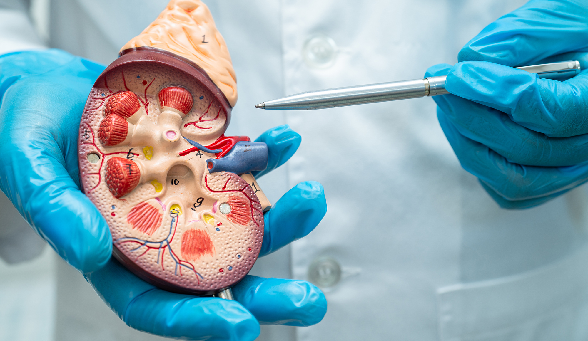
Intravesical immunotherapy using Bacillus Calmette-Guérin (BCG) is a localised treatment for non-muscle invasive bladder cancer (NMIBC). This approach involves the direct introduction of live, weakened bacteria into the bladder using a catheter.

BCG activates the body’s immune system to target and destroy bladder cancer cells, helping to prevent the recurrence and progression of the disease. It is considered an effective form of immunotherapy that operates primarily within the bladder, minimising systemic side effects.

Indications for Intravesical Immunotherapy with BCG

Intravesical immunotherapy with BCG is specifically indicated for several scenarios in the treatment of bladder cancer:

  • Non-Muscle Invasive Bladder Cancer (NMIBC): BCG is primarily used for NMIBC, particularly for high-risk cases where there is a greater likelihood of recurrence or progression.
  • After TURBT: It is commonly administered after transurethral resection of a bladder tumour (TURBT) to reduce the risk of cancer recurrence.
  • Carcinoma in Situ (CIS): This high-risk form of NMIBC, which is flat and often difficult to detect, responds well to BCG therapy.
  • Prophylactic Treatment: BCG can be used as a preventive treatment to delay or prevent the recurrence of bladder cancer following surgery.

Preparation for BCG Treatment

Proper preparation is essential to ensure the safety and effectiveness of intravesical immunotherapy with BCG. Here are the necessary steps patients typically follow before undergoing this treatment:

Medical Evaluation

Patients undergo a comprehensive medical evaluation to confirm the suitability of BCG therapy. This includes reviewing their medical history, current health status, and any previous treatments for bladder cancer.

Laboratory Tests

Blood tests, urine analysis, and possibly urine cultures are performed to check for underlying conditions that might affect the treatment.

Bladder Examination

A thorough bladder examination, often through cystoscopy, is conducted to ensure no active tumours or infections.

Medication Review

Patients may need to adjust or temporarily stop certain medications, especially those that could interfere with immune response or increase the risk of bleeding.

The BCG Treatment Process

The process of administering BCG treatment for bladder cancer is meticulously planned to maximise therapeutic effectiveness while minimising discomfort. Here is an outline of the typical procedure:

Catheter Insertion

The treatment begins with inserting a catheter into the bladder through the urethra. This procedure is usually done in a hospital or clinic under sterile conditions.
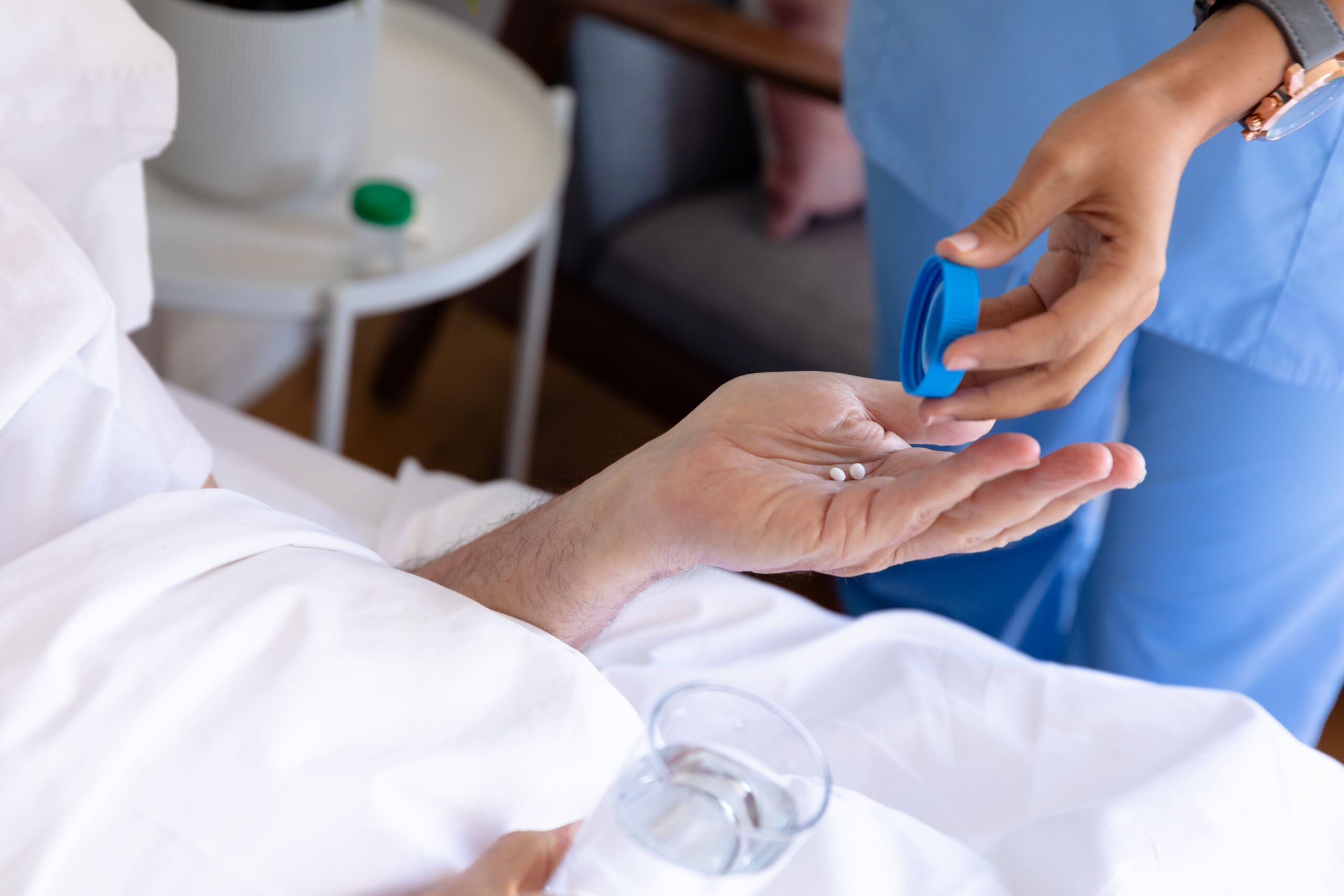
BCG Instillation

Once the catheter is in place, the BCG solution, which contains a live attenuated strain of the tuberculosis bacterium, is instilled into the bladder. The catheter is removed, allowing the solution to remain in the bladder.

Retention Time

Patients are usually asked to hold the solution in their bladder for about one to two hours. This retention time is crucial as it allows the BCG to come into direct contact with the bladder wall, where it exerts its immunotherapeutic effects.

Post-Instillation Instructions

After the retention period, patients are advised to urinate in order to expel the BCG solution. To minimise the risk of spreading the bacteria, it is recommended to use a bleach solution to disinfect the toilet after each use for the first few hours post-treatment.

Treatment Schedule

BCG therapy is typically given once a week for six weeks. Depending on the patient’s response, this initial course is followed by additional maintenance treatments.

Post Treatment Care and Recovery

After completing a session of BCG treatment, proper care is crucial to manage side effects and ensure the best therapeutic outcomes. Here are the key aspects of post-treatment care and recovery:

  • Hydration: Patients are encouraged to drink plenty of fluids after the treatment to help flush the bladder and reduce the concentration of BCG in the urine.
  • Pain Management: Over-the-counter pain relievers may address discomfort or mild pain from the catheter or the BCG solution.
  • Monitoring for Side Effects: Common side effects include urinary frequency, discomfort during urination, and flu-like symptoms such as fever and fatigue. Persistent or severe symptoms should be reported.
  • Follow-Up Visits: Regular follow-up appointments are essential to monitor the effectiveness of the treatment and manage any side effects. These visits may include urine tests, cystoscopy, and discussions about symptom management.
  • Precautions: Patients are advised to avoid sexual activity for 48 hours after each treatment and may be instructed to use a condom for several weeks post-treatment to protect their partners, as BCG is live bacteria.

Risks and Complications

While BCG immunotherapy is generally safe and effective for treating non-muscle invasive bladder cancer, it can have potential risks and complications, which are important for patients to be aware of:

  • Infection: Although rare, live bacteria in the bladder can lead to infection. Symptoms may include persistent fever, chills, and worsening urinary symptoms.
  • BCG Reaction: Some patients may experience a severe reaction to BCG, characterised by intense pain, frequent urination, blood in the urine, or a prolonged high fever. This requires immediate medical attention.
  • Bladder Irritation: Common side effects include cystitis-like symptoms such as urgency, frequency, and dysuria (painful urination). These symptoms usually resolve within a few days but can be uncomfortable.
  • Systemic BCGosis: Very rarely, the BCG can spread beyond the bladder, leading to a systemic infection. This serious complication is more likely in patients with compromised immune systems and necessitates urgent treatment.
  • Contracting Tuberculosis: There is a minimal risk of contracting tuberculosis from the BCG strain; however, it is a possibility that requires vigilant monitoring.

Conclusion

Patients considering BCG immunotherapy should be aware of its potential side effects and the importance of adherence to post-treatment guidelines. Continuous monitoring and proper management of symptoms post-treatment can significantly enhance the effectiveness and safety of this therapy.

Interstitial Cystitis Treatment Options: A Guide For Prospective Patients

Interstitial cystitis, often referred to as painful bladder syndrome, is a chronic condition that causes bladder pressure, bladder pain, and sometimes pelvic pain. The symptoms can vary greatly between people and even in the same person over time.

Since there’s no cure yet, treatment focuses on easing these symptoms. By exploring various treatment options, those affected can take active steps in managing their condition and making choices that best fit their health needs.

What is Interstitial Cystitis?

Interstitial cystitis (IC) is a chronic condition that affects the bladder and pelvic area, often resulting in discomfort and pain. Its symptoms and causes are varied and unclear, making diagnosis and treatment challenging.

Symptoms of Interstitial Cystitis

The most common symptoms include:

  • Chronic Pelvic Pain: Worsens as the bladder fills and may decrease after urination.
  • Increased Urinary Frequency: Frequent urination during both day and night.
  • Urgency: A persistent, urgent need to urinate.
  • Pain During Sexual Intercourse: Discomfort or pain during sexual activities.

Causes of Interstitial Cystitis

The causes of IC are not fully understood, which complicates treatment approaches. Some possible causes include:

  • Defective Bladder Lining: Potential damage to the bladder lining, allowing urine to irritate the bladder wall.
  • Autoimmune Response: An autoimmune reaction may be involved, targeting the bladder.
  • Possible Infection: An infectious cause has been considered but not proven.
  • Systemic Conditions: IC might be related to broader systemic issues.

Interstitial Cystitis Dietary Modifications

Let’s delve into how dietary choices can impact interstitial cystitis (IC). Certain foods and beverages have been observed to exacerbate IC symptoms. Through the identification and avoidance of these triggers, patients can effectively alleviate their discomfort.

Common Dietary Irritants

  • Acidic Foods: Citrus fruits, tomatoes, and vinegar-based products can irritate the bladder.
  • Spicy Foods: Items containing chillies or other strong spices may provoke symptoms.
  • Caffeinated Beverages: Coffee, tea, and some sodas can increase urinary frequency and urgency.
  • Alcoholic Drinks: Alcohol is a known bladder irritant and can worsen symptoms.

Beneficial Foods

  • Alkaline Foods: Non-citrus fruits, vegetables, and whole grains help neutralise bladder acidity.
  • Water: Increasing water intake can dilute urine, reducing bladder wall irritation.

Tips for Dietary Management

  • Food Diary: Keeping a diary to track foods and symptoms can help identify personal triggers.
  • Balanced Diet: Ensuring a nutritionally balanced diet is important even while eliminating irritants.

Oral Medications

Oral medications are a common and effective treatment option for managing the symptoms of interstitial cystitis. They work by various mechanisms to reduce discomfort, frequency of urination, and bladder inflammation.

Antihistamines

Medications such as hydroxyzine can help reduce urinary urgency and frequency by counteracting the effects of histamine, which is involved in inflammation.

Tricyclic Antidepressants

Amitriptyline is used to help relax the bladder and block pain signals. It is effective in reducing both pain and urinary frequency.

Physical Therapy and Pain Management

Physical therapy and pain management strategies are integral to treating interstitial cystitis, especially for alleviating pelvic pain and improving pelvic floor function. These approaches include:

Physical Therapy Techniques

  • Pelvic Floor Therapy: Specialised physical therapists use techniques to relax tight pelvic floor muscles, which are often a source of pain and urinary issues in IC patients.
  • Manual Therapy: Techniques such as soft tissue mobilisation can help decrease muscle tenderness and pelvic discomfort.

Pain Management Strategies

  • Heat Therapy: Applying heat to the pelvic area can soothe muscle pain and abdominal discomfort.
  • TENS (Transcutaneous Electrical Nerve Stimulation): This method uses electrical impulses to reduce pain by blocking nerve signals to the brain.
  • Pain Medication: Non-opioid pain relievers, such as acetaminophen or non-steroidal anti-inflammatory drugs (NSAIDs), are recommended to manage chronic pain, with careful consideration of their effects on bladder irritation.

Intravesical Treatments

Intravesical treatments involve instilling medication directly into the bladder via a catheter to target interstitial cystitis symptoms at their source. This method is often pursued when other options have proven inadequate.

Treatments typically occur weekly for six to eight weeks and are conducted in a doctor’s office. Eligibility and treatment frequency are determined based on their specific symptoms and response to other therapies.

Common Intravesical Solutions

  • Dimethyl Sulfoxide (DMSO): The only FDA-approved intravesical solution for IC, DMSO helps reduce inflammation and pain and has a muscle-relaxing effect on the bladder.
  • Heparin: Similar to the bladder’s natural lining, heparin may help restore the bladder surface and prevent irritation from urine.
  • Lidocaine: An anaesthetic used to numb the bladder wall, reducing pain and urgency.

Surgical Options

Surgical interventions for interstitial cystitis are typically considered as a last resort when all other treatments have failed to provide adequate relief. These procedures aim to alleviate symptoms by addressing the bladder directly.

  • Bladder Distension: A procedure where the bladder is filled with fluid to stretch its walls, potentially reducing pain signals.
  • Bladder Fulguration: Involves using electricity or a laser to burn away ulcers and areas of inflammation inside the bladder.
  • Resection of Hunner’s Ulcers: Specific ulcers, known as Hunner’s ulcers, are surgically removed to decrease bladder pain and irritation.
  • Bladder Augmentation: A major surgical procedure that increases the bladder capacity by adding a piece of the intestine to the bladder wall.
  • Urinary Diversion: In severe cases, the normal urine flow is diverted from the bladder to an external pouch, significantly reducing or eliminating symptoms.

Conclusion

It’s evident that each treatment option presented in this blog holds promise for alleviating symptoms and enhancing daily functioning. The optimal choice hinges on the person’s specific needs and the severity of their symptoms. By considering these factors thoughtfully, prospective patients can work with their doctor to devise a personalised treatment plan that offers the greatest potential for relief and improved quality of life.

Recovery After Kidney Stone Removal: A Urologist’s Guide

Kidney stones are hard mineral and salt deposits that form in the kidneys. They can cause significant pain and discomfort, often requiring medical intervention for removal. The process of recovering from kidney stone removal is important to ensure the health and well-being of the patient.

Kidney Stone Removal Procedures

Kidney stone removal can be performed using various medical procedures, depending on the size, location, and type of the stones. Common methods include:

Extracorporeal Shock Wave Lithotripsy (ESWL)

ESWL involves using shock waves to break the kidney stones into smaller pieces that can be passed naturally through the urinary tract. It is a non-invasive procedure typically used for stones less than 2 centimetres in diameter.

Ureteroscopy

This procedure involves inserting a small scope through the urethra and bladder to reach the ureters or kidneys. Small stones are removed or broken down using a laser or mechanical device.

Percutaneous Nephrolithotomy

For larger or irregularly shaped stones, percutaneous nephrolithotomy may be required. This involves making a small incision in the back to insert instruments directly into the kidney, allowing for the removal of large stones.

Each of these procedures has specific indications, and the choice of method depends on various factors, including the patient’s overall health, the characteristics of the kidney stones, and the presence of any complicating conditions. Understanding the procedure used is the first step in managing expectations and planning for a smooth recovery.

Immediate Post-Operative Care

Following the removal of kidney stones, immediate post-operative care is critical to ensure a smooth and safe recovery. Key aspects of this care include:

  • Monitoring: Patients are closely monitored for signs of complications, including bleeding, infection, or adverse reactions to anaesthesia. Vital signs, fluid balance, and pain levels are regularly checked during the initial recovery phase in the hospital.
  • Hydration: Increased fluid intake is important after kidney stone removal to help flush out any remaining stone fragments and prevent new stones from forming. Patients are encouraged to drink at least 2 to 3 litres of water per day unless otherwise advised by their urologist.
  • Pain Relief: Pain management is a priority, and medications are often prescribed to alleviate discomfort after the procedure. The type and intensity of pain relief required can vary depending on the specifics of the surgery and the person’s pain tolerance.
  • Stent Placement: Occasionally, a temporary ureteral stent is placed to keep the ureter open, allowing urine to flow freely. This can help prevent blockages from swelling or remaining stone fragments. The stent is usually removed during a simple outpatient procedure after a few weeks.
  • Rest: Patients are advised to rest and limit physical activity for several days post-surgery to allow the body to heal. Specific guidelines on activity levels will be provided by the urologist based on the patient’s health status and the type of procedure performed.

Pain Management Strategies

Effective pain management is a critical component of the recovery process after kidney stone removal. Here are some strategies commonly recommended by urologists:

  • Medication: Depending on the intensity of the pain and the patient’s overall health, various pain relief medications may be prescribed. These can include over-the-counter pain relievers such as acetaminophen or ibuprofen, and in cases of more severe pain, stronger prescription opioids may be temporarily used.
  • Heat Therapy: Applying heat can help soothe the area and reduce discomfort. A heating pad or a warm cloth applied to the back or abdomen may ease muscle spasms and alleviate pain.
  • Activity Modification: Limiting strenuous activities and ensuring adequate rest can help manage pain during the initial recovery period. Gradual reintroduction of normal activities is advised as pain subsides and healing progresses.
  • Follow-Up Care: Regular follow-up with the urologist is important for adjusting pain management strategies as the recovery progresses. This ensures that any persistent or unusual pain can be addressed promptly.

Diet and Nutrition Recommendations

Diet plays a pivotal role in the recovery and prevention of future kidney stones. Here are key dietary guidelines to follow after kidney stone removal:

  • Calcium-Rich Foods: Contrary to popular belief, consuming calcium-rich foods does not necessarily contribute to calcium oxalate stones; dietary calcium can help prevent their formation by binding with oxalate in the intestines.
  • Reduce Sodium Intake: High sodium intake can increase calcium buildup in urine, possibly leading to new stones. Limit salt consumption to help manage this risk.
  • Limit Oxalate-Rich Foods: If the removed stones were calcium oxalate, reducing the intake of oxalate-rich foods (such as spinach, rhubarb, and almonds) can help prevent recurrence.
  • Protein Intake: Moderate consumption of animal protein is advised, as excessive intake can increase the risk of stone formation.

Physical Activity and Rest

Balancing physical activity and rest is essential for effective recovery after kidney stone removal. Here are some guidelines to follow:

  • Gradual Resumption of Activities: Patients should gradually increase their activity level as their urologist recommends. Starting with light activities such as walking can help stimulate recovery without overstressing the body.
  • Avoid Strenuous Activities: For several weeks post-surgery, it is advisable to avoid heavy lifting and high-impact activities that could strain the surgical area or dislodge any healing tissues.
  • Adequate Rest: Ensuring sufficient rest is important during the initial recovery phase. Rest helps the body heal and reduces the risk of complications.
  • Sleep Quality: Maintaining good sleep hygiene can enhance recovery. Aim for 7-8 hours of sleep daily to support overall health and well-being.
  • Listen to Your Body: Patients should pay close attention to their body’s signals. If pain increases or new symptoms appear during physical activity, they should decrease the intensity or consult their urologist.

By following these recommendations, patients can ensure a balanced recovery, minimising the risk of complications and promoting faster healing.

Monitoring and Managing Potential Complications

Effective monitoring and management of potential complications are important in the post-operative recovery from kidney stone removal. Here are some common complications to be aware of and strategies for managing them:

  • Infection: Signs of infection may include fever, chills, or unusual pain. Prompt treatment with antibiotics, as prescribed by a urologist, is essential to manage any infections that arise.
  • Bleeding: Minor bleeding might be normal, but excessive or prolonged bleeding should be reported to a urologist immediately.
  • Urinary Retention: Difficulty in urinating can occur, especially if a stent is placed. Drinking plenty of fluids and sometimes medications can help alleviate this issue.
  • Stent Discomfort: Patients might experience discomfort or a sensation of urgency if a stent is in place. Medications can be prescribed to ease these symptoms.
  • Stone Fragments: Small stone fragments may pass after the procedure, causing pain or irritation. Maintaining high fluid intake helps flush these fragments out of the urinary system.

Long-Term Health Management

Long-term health management after kidney stone removal is vital to prevent recurrence and maintain overall urinary tract health. Here are strategies to ensure effective long-term management:

  • Regular Health Check-ups: Routine visits to a urologist are important to monitor kidney health and detect any signs of new stone formation early.
  • Continued Hydration: Maintaining a high fluid intake, especially water, should be a lifelong habit to prevent the formation of new stones.
  • Dietary Adjustments: Adhering to the dietary recommendations provided earlier, such as reducing sodium and oxalate intake and balancing calcium, helps minimise the risk of future stones.
  • Medication Compliance: If medications like potassium citrate are prescribed to prevent stone formation, it is important to follow the prescribed regimen closely. The type and necessity of medication can vary based on the person’s health conditions and specific medical advice.
  • Lifestyle Modifications: Incorporating regular exercise, maintaining a healthy weight, and avoiding smoking can also help reduce the risk of kidney stones.

By committing to these long-term health strategies, patients can significantly lower their chances of recurrent kidney stone issues and enhance their overall health.

Conclusion

Recovering from kidney stone removal involves careful attention to immediate post-operative care, effective pain management, and adherence to dietary and physical activity guidelines. Monitoring for potential complications and engaging in long-term health management is important to preventing recurrence and maintaining overall urinary health.

Patients need to maintain open communication with their urologist and adhere to recommended practices to ensure the best possible outcomes in their recovery journey.

Kidney Stone Pain Locations: A Urologist’s Guide

Kidney stones, also known as renal calculi, are solid masses made of crystals. They originate in the kidneys but can develop anywhere along the urinary tract, which includes the kidneys, ureters, bladder, and urethra. Several factors, including diet, excess body weight, some medical conditions, and certain supplements and medications, influence the formation of kidney stones.

The symptoms of kidney stones can be acute and include severe pain, hematuria (blood in the urine), nausea, vomiting, and sometimes fever if an infection is present. The pain is often described as one of the most severe types of pain and can be a key indicator of the stone’s presence and movement within the urinary tract.

Anatomy of the Kidney and Urinary Tract

The kidneys, bean-shaped organs located below the rib cage on each side of the spine, are important for filtering waste and excess substances from the blood to produce urine. Each kidney connects to a ureter, a narrow tube that transfers urine to the bladder, where it is stored until expelled during urination through the urethra.

This pathway is critical not only for waste removal and fluid balance but also for facilitating the movement of kidney stones. The pain associated with kidney stones often occurs as these stones move through the narrow ureters, highlighting the importance of this anatomy in understanding pain locations.

Common Pain Locations Associated with Kidney Stones

The pain caused by kidney stones, known medically as renal colic, is typically very sharp, intermittent, and can be extremely intense. Common pain locations include:

  • Flank Pain: The most common site of pain is in the flank, just below the ribs on the back, where the kidneys are located. This pain can radiate to the lower abdomen and groin as the stone moves down the urinary tract.
  • Side and Back Pain: Pain can also be felt along the side and back, directly above the hip bone.
  • Lower Abdomen and Groin Discomfort: As the stone approaches the bladder, pain may shift towards the lower abdomen and groin area. Men may also experience testicular pain, while women might feel pain in the labial area.

While the locations of pain described are typical for kidney stones, it is important to note that the intensity and specific locations of pain can vary significantly from person to person.

Factors Influencing Pain Location

The location and intensity of pain caused by kidney stones can vary significantly depending on several factors:

  • Size of the Stone: Larger stones are more likely to cause pain as they move through the urinary tract because they can cause more significant obstruction. Smaller stones, while sometimes passing more easily, can still cause sharp pain due to their movement.
  • Location of the Stone: Stones located in the kidney typically cause pain in the flank area. As the stone moves into the ureter, the pain can shift towards the abdomen and groin.
  • Shape of the Stone: Jagged or irregularly shaped stones can cause more irritation to the urinary tract lining, potentially increasing pain levels as they travel towards the bladder.
  • Ureter Narrow Passages: Narrower sections of the ureter can cause stones to become lodged, intensifying pain as the body tries to pass the stone through these narrow areas.

Understanding these factors is important for medical professionals to predict pain patterns and provide appropriate interventions to manage pain effectively.

Diagnosing Kidney Stones

Diagnosing kidney stones typically involves a combination of clinical assessment and diagnostic imaging. The process often includes:

  • Patient History and Symptoms: A detailed discussion about the patient’s symptoms, pain locations, and medical history helps guide further diagnostic steps.
  • Physical Examination: A physical exam can help identify the pain areas and assess for signs related to kidney stones.
  • Urinalysis: This test checks for blood, bacteria, and crystals in the urine, as well as indicators of kidney stones or an associated infection.
  • Blood Tests: Blood tests can evaluate kidney function and detect signs of infection or other related abnormalities.
  • Imaging Studies: The most definitive tools for diagnosing kidney stones are imaging studies. Common methods include:
    • Ultrasound: A non-invasive test that can detect stones and assess kidney swelling.
    • CT Scan: Highly effective at identifying kidney stones and providing detailed images of the kidney, ureters, and bladder.

Management and Treatment Options

The management and treatment of kidney stones depend on the size, location, and type of stone, as well as the patient’s symptoms. Treatment options include:

  • Medical Therapy: For smaller stones, patients may be advised to drink plenty of fluids to help pass the stone naturally. Pain relief medications are commonly prescribed during this process.
  • Medical Expulsive Therapy (MET): This involves the use of medications such as alpha-blockers, which can help relax the muscles in the ureter, allowing the stone to pass more easily.
  • Extracorporeal Shock Wave Lithotripsy (ESWL): ESWL uses shock waves to break larger stones into smaller pieces that can be passed naturally in the urine. Although considered non-invasive, ESWL can sometimes cause side effects such as bruising and discomfort; thus, it might be perceived as minimally invasive. It remains one of the most frequently used methods to treat kidney stones due to its effectiveness for certain stone types.
  • Ureteroscopy: For stones in the ureter, a ureteroscope is inserted through the urethra to break up or remove the stone. Although ureteroscopy is a common method for removing stones lodged in the ureter, the procedure can vary in complexity and sometimes involves risks such as infection, bleeding, or injury to the ureter.
  • Percutaneous Nephrolithotomy: This surgical procedure is used for very large stones or stones that cannot be treated with less invasive methods. It involves making a small incision in the back to access the kidney directly and remove the stones.

Conclusion

Kidney stones are a common and often intensely painful condition that can affect anyone. Understanding the locations and nature of the pain, along with the factors influencing these aspects, helps in a timely and accurate diagnosis.

Early detection and management are important to alleviate symptoms and prevent complications. With a variety of treatment options available, most people can effectively manage and treat kidney stones, often without needing surgical intervention.

Kidney Stone Treatment Options: A Guide For Prospective Patients

Kidney stones, or renal lithiasis, are hard deposits of minerals and salts that form inside the kidneys, typically resulting from the concentration of minerals in the urine. These stones can be composed of various substances such as calcium oxalate, uric acid, struvite, or cystine, depending on dietary habits, fluid intake, and genetic predisposition.

Symptoms often do not appear until the stone moves within the kidney or into the ureter, causing severe pain, nausea, vomiting, and blood in the urine. Without appropriate treatment, kidney stones can lead to significant complications, including infections and kidney damage.

Factors Influencing the Recommendation of Kidney Stone Treatment

The decision to treat kidney stones is influenced by several key factors, each determining the necessity and urgency of medical intervention:

  • Size and Location: Treatment is typically necessary for stones larger than 5 millimetres, as they may not pass naturally and are more likely to cause obstruction in the urinary tract. Smaller stones, less than 5 millimetres in diameter, might pass on their own, though medical monitoring and advice are recommended to manage pain and monitor for potential complications.
  • Symptoms and Complications: Severe pain and symptoms of urinary tract infections are strong indicators for treatment. Bleeding in the urine, a common symptom of kidney stones, also requires evaluation to rule out potential kidney damage.
  • Risk Factors and Medical History: Patients with a history of kidney stones, especially those with underlying conditions like hyperparathyroidism or metabolic disorders that predispose them to recurrent stone formation, often require proactive treatment to manage the condition and prevent new stones from forming.

What is The Diagnosis for Kidney Stones?

Diagnosing kidney stones typically involves a combination of physical examination, review of symptoms, and diagnostic imaging. The process is aimed at confirming the presence of stones, assessing their size and location, and determining the appropriate treatment plan.

  • Medical History and Symptom Review: The initial diagnostic step includes a detailed discussion of the patient’s symptoms, medical history, and any previous incidents of kidney stones. This helps to assess risk factors such as dietary habits, fluid intake, and family history of kidney stones.
  • Physical Examination: A physical exam may help identify pain in the kidney area. Urologists also look for signs of fever and infection, which could indicate complications from a kidney stone.
  • Urinalysis: Testing the urine can reveal crystals, bacteria (indicating infection), or blood, all of which suggest the presence of a stone.
  • Blood Tests: Blood work helps evaluate kidney function and measure levels of substances that could promote stone formation, such as calcium, uric acid, and creatinine.
  • Diagnostic Imaging: Imaging tests are important in the diagnosis of kidney stones. Common methods include:
    • Ultrasound: A non-invasive test ideal for detecting stones and assessing the urinary tract, while CT scans provide detailed images, making them highly effective in identifying even small stones.
    • X-rays: Often used to locate stones that are visible using this technique.
    • CT Scan: Provides detailed images and is highly effective in detecting even small stones within the urinary tract.

Together, these diagnostic tools provide a comprehensive view of the urinary system, enabling precise identification of kidney stones and aiding in the formulation of a targeted treatment strategy.

Preparing for Kidney Stone Treatment

Preparation for kidney stone treatment involves several steps to optimise patient safety and treatment outcomes. These preparations vary depending on the type of treatment planned, but common measures include:

  • Medical Evaluation: Prior to any treatment, a comprehensive medical evaluation is necessary to assess the patient’s overall health and suitability for the chosen treatment method. This includes evaluating kidney function and checking for any other medical conditions that might complicate treatment.
  • Dietary and Fluid Adjustments: Patients may be advised to adjust their diet or increase fluid intake before treatment to help manage the size and composition of kidney stones.
  • Medications: Some treatments may require patients to stop or adjust certain medications temporarily, especially those that affect blood clotting or other relevant physiological processes.

Treatment Options for Kidney Stones

Treatment for kidney stones varies depending on the size, type, and location of the stones, as well as the patient’s overall health and symptoms. Options range from conservative management to more invasive procedures:

Conservative Management

Small stones that are likely to pass naturally may only require pain management and increased fluid intake to facilitate stone passage. Continuous monitoring ensures stones pass without complications.

Medical Therapy

Certain medications can help manage symptoms or aid in the passage of kidney stones. These include pain relievers, alpha-blockers to relax the muscles in the ureter, and medications to control nausea.

Extracorporeal Shock Wave Lithotripsy (ESWL)

This non-invasive treatment uses shock waves to break stones into smaller pieces that can be passed more easily in the urine.

Ureteroscopy

For stones lodged in the ureter or kidney, a small scope is passed through the bladder into the ureter. Stones are either removed or broken up using laser energy.

Percutaneous Nephrolithotomy

For larger or more complex stones, a small incision is made in the back to allow direct removal of the stone from the kidney.

Open Surgery

Although rarely necessary due to advances in less invasive techniques, open surgery may be required in very complex cases where other treatments are not suitable.

Each treatment option has specific risks and benefits, with the choice depending on the person’s circumstances and stone characteristics.

Post-Procedure Care and Recovery

The recovery process following kidney stone treatment varies based on the type of procedure performed and the patient’s health. Effective post-procedure care is critical for ensuring a smooth recovery and preventing further complications or recurrence of kidney stones:

  • Pain Management: Discomfort is common after kidney stone removal procedures, especially following shock wave lithotripsy or more invasive surgeries. Effective pain management typically involves prescribed pain relievers or over-the-counter medications.
  • Fluid Intake: Increasing fluid intake post-procedure is important to help flush out any remaining stone fragments and prevent new stones from forming. Patients are often advised to drink at least two to three litres of water daily.
  • Activity Restrictions: Patients may need to limit physical activity, especially following invasive procedures, to allow the body to heal. The duration of activity restriction can vary depending on the specific treatment.
  • Follow-Up Appointments: Regular follow-up appointments are essential to monitor recovery, check for the presence of remaining stone fragments, and manage any complications. Imaging tests may be repeated to ensure that all stone fragments have been cleared.
  • Dietary Modifications: Depending on the type of stones, a urologist may recommend dietary changes to prevent recurrence. This might include reducing intake of salt, protein, or certain foods high in oxalates or calcium.

Adhering to these guidelines helps minimise recovery time, reduces the risk of complications, and decreases the likelihood of future kidney stones.

Conclusion

Effective treatment and management of kidney stones require a comprehensive approach that includes accurate diagnosis, tailored medical interventions, and diligent post-procedure care. Advances in medical technology have significantly enhanced the safety and efficacy of treatments, improving outcomes for patients. By adhering to medical advice and making necessary lifestyle changes, patients can effectively manage their condition and reduce the risk of recurrence, ultimately improving their quality of life.

Partial Nephrectomy: A Urologist’s Guide

Partial nephrectomy, also known as nephron-sparing surgery, primarily focuses on removing a kidney tumour while preserving as much surrounding healthy kidney tissue as possible.

This procedure is especially beneficial for maintaining overall kidney function and is preferred over radical nephrectomy, which involves removing the entire kidney. By preserving kidney tissue, partial nephrectomy minimises the risk of chronic kidney disease after surgery. Read on to learn more about partial nephrectomy, including the surgical techniques and postoperative care involved.

When is Partial Nephrectomy Recommended?

Partial nephrectomy is recommended for people with localised kidney tumours when preserving renal function is a priority. It is most commonly recommended in the following scenarios:

  • Small Renal Masses: Typically, tumours that are 4 cm or smaller (T1a) are ideal candidates for partial nephrectomy. These small masses are often detected incidentally during imaging for other conditions.
  • Solitary Kidney: For patients with only one functioning kidney, partial nephrectomy is important for preserving as much renal function as possible, thus avoiding the need for dialysis.
  • Bilateral Renal Tumours: When tumours are present in both kidneys, partial nephrectomy can be performed to treat tumours on both sides, thus maintaining some degree of kidney function.
  • Pre-existing Renal Conditions: In individuals with pre-existing conditions such as chronic kidney disease, where overall renal function is already compromised, nephron-sparing surgery helps to preserve the remaining renal function.
  • Genetic Predisposition: Patients with hereditary syndromes that predispose them to multiple kidney tumours, such as von Hippel-Lindau disease, are also candidates for multiple partial nephrectomies over their lifetime.

Preoperative Preparation and Assessment

The preoperative preparation and assessment phase includes a comprehensive evaluation of the patient’s overall health and kidney function.

  • Health Assessment: A thorough medical evaluation is conducted to assess the patient’s general health and identify any coexisting medical conditions that may affect surgery or anaesthesia.
  • Renal Function Tests: Kidney function is assessed through blood tests, including serum creatinine and blood urea nitrogen (BUN) levels, to ensure the remaining kidney can function adequately post-surgery.
  • Imaging Studies: Techniques such as computed tomography (CT) scans, magnetic resonance imaging (MRI), or ultrasound are used to determine the tumour’s size, location, and relationship to surrounding structures in the kidney.
  • Renal Artery Imaging: Specialised imaging techniques, such as CT angiography or MR angiography, provide a detailed view of the renal vasculature. This is important for surgical planning to minimise blood loss and ensure precise tumour removal.

Surgical Techniques

The surgical techniques for partial nephrectomy can vary based on the tumour’s characteristics and the patient’s overall health. Here are the primary approaches used:

Open Partial Nephrectomy

This traditional method involves a larger incision to access the kidney directly. It is often used for complex tumours or when the surgeon requires more direct visualisation and access.

Laparoscopic Partial Nephrectomy

This minimally invasive approach uses small incisions and a camera to guide the surgery. This method typically results in less pain and a quicker recovery compared to open surgery.

Robot-Assisted Laparoscopic Partial Nephrectomy

This is an advanced form of laparoscopic surgery where the surgeon uses a robotic system to enhance precision, flexibility, and control during the operation. This technique is beneficial for intricate surgeries.

Key Surgical Considerations

  • Ischaemia Time: This refers to when blood flow to the kidney is reduced or halted to facilitate tumour removal. Minimising ischaemia time is crucial to protect kidney function.
  • Tumour Location: The approach may vary significantly depending on whether the tumour is located near the surface or deep within the kidney.
  • Tumour Size and Complexity: Larger or more complex tumours might require more extensive reconstruction of the kidney post-tumour removal.

Postoperative Care and Management

Effective postoperative care is essential for a successful recovery after a partial nephrectomy.

  • Monitoring: Regular assessment of kidney function through blood tests and urine output is critical to ensure the remaining kidney compensates adequately.
  • Pain Management: Pain is managed through medications, which may include oral or intravenous options, depending on the intensity of the pain and the patient’s overall health.
  • Infection Prevention: Prophylactic antibiotics may be administered to prevent infections, and wound care is meticulously managed to ensure clean, dry, and intact dressings.
  • Physical Activity: A gradual increase in physical activity is encouraged to promote circulation and prevent blood clots. Patients are typically advised to avoid strenuous activities or heavy lifting for several weeks.
  • Dietary Management: Adequate hydration and a balanced diet rich in essential nutrients support healing and kidney health.

Conclusion

Partial nephrectomy is a crucial surgical option for individuals diagnosed with kidney tumours, designed to remove the malignancy while preserving as much kidney functionality as possible. This approach aims to benefit patients by supporting better long-term kidney health and overall well-being.

If you’re considering partial nephrectomy or seeking more information about your options, get in touch with us. Our clinic provides expert consultations and treatment plans tailored to meet personalised patient needs.

Urinary Tract Infection (UTI) Prevention Guide: What You Need To Know

Urinary tract infections (UTIs) are commonly associated with women but also represent a significant concern for men. UTIs in men may affect any component of the urinary system, including the kidneys, ureters, bladder, and urethra. Typically caused by bacteria, E. coli (Escherichia coli) is the most frequent culprit.

Although UTIs are relatively uncommon in younger men and may suggest an underlying anatomical or functional issue, the risk increases with age in older men due to factors such as prostate enlargement, which impedes urinary flow.

What Are the Common Causes and Risk Factors?

The primary cause of UTIs in men is the invasion of the urinary tract by bacteria, predominantly Escherichia coli, which normally resides in the bowel. Other bacteria such as Klebsiella pneumoniae and Proteus mirabilis can also cause these infections.

The following are some key risk factors that increase the likelihood of developing UTIs:

Anatomical Factors

  • Enlarged Prostate: An enlarged prostate gland can restrict urine flow, thereby increasing the risk of infection.
  • Urethral Stricture: Scar tissue-induced narrowing of the urethra can impede urine flow and increase UTI risk.

Medical Conditions

  • Diabetes: Men with diabetes are at higher risk due to the immune system’s compromised ability to fight off infections.
  • Neurological Conditions: Conditions that impair bladder function or urinary retention can contribute to the risk of UTIs.

Lifestyle and Behavioral Factors

  • Poor Hygiene: Inadequate genital hygiene can increase the risk of bacterial spread to the urinary tract.
  • Sexual Activity: Sexually active men, especially those with multiple partners, may have a higher risk of UTIs due to the potential introduction of bacteria during intercourse.

What Are the Symptoms of UTIs in Men?

The symptoms of urinary tract infections in men can vary depending on the location of the infection within the urinary tract. Common symptoms include:

Lower Urinary Tract Symptoms

  • Dysuria: Pain or burning sensation during urination.
  • Increased Frequency of Urination: An urgent need to urinate more frequently than usual, even when the bladder is not full.
  • Hesitancy and Dribbling: Difficulty starting urination and a weak or interrupted urine stream.
  • Nocturia: Increased frequency of urination during the night.

Upper Urinary Tract Symptoms

  • Flank Pain: Pain or discomfort in the lower back or sides, which can indicate a kidney infection, also known as pyelonephritis.
  • Fever and Chills: These symptoms may occur if the infection has spread to the kidneys, suggesting a more serious condition.

General Symptoms

  • Cloudy or Bloody Urine: The appearance of urine may change, becoming cloudy or red, which is a sign of blood in the urine.
  • Unpleasant Odour: Urine may have a strong or foul smell.

Key Prevention Strategies for UTIs

Effective prevention of UTIs can significantly reduce the incidence and severity of these infections.

Hydration and Diet

  • Adequate Fluid Intake: Regular consumption of fluids helps flush out bacteria from the urinary tract.
  • Balanced Diet: Maintaining a diet rich in fruits, vegetables, and fibre can help minimise the risk of UTIs.

Personal Hygiene

  • Proper Genital Hygiene: Men should wash the genital area daily and after sexual intercourse to reduce the risk of bacterial transmission.
  • Regular Urination: It is advisable to urinate frequently to prevent bacterial accumulation in the urinary tract.
  • Post-Intercourse Urination: Urinating immediately after sexual activity can help expel any bacteria that may have entered the urethra.

Medical and Natural Interventions

  • Probiotics: Consuming probiotics can support beneficial bacteria levels in the gut and around the genital area, potentially reducing UTI risk.

Lifestyle Adjustments

  • Avoiding Irritants: Reducing the intake of bladder irritants such as caffeine, alcohol, and acidic foods can help maintain a healthy urinary tract.
  • Suitable Clothing: Opting for loose, cotton underwear helps keep the genital area dry, decreasing the likelihood of bacterial growth.

By incorporating these preventive strategies into daily routines, men can effectively reduce their risk of developing urinary tract infections.

When to Seek Medical Attention

UTIs can progress and potentially lead to more serious complications if not treated promptly. Here are guidelines on when you should consult a healthcare provider:

  • Persistent Symptoms: If symptoms such as painful urination, frequent urination, urgency to urinate, or lower abdominal pain persist despite home remedies and preventive measures.
  • Changes in Urine Appearance: Cloudy, bloody, or unusually strong-smelling urine that does not improve.
  • Flank Pain: Pain or tenderness in the back or sides can indicate a kidney infection requiring immediate medical attention.
  • Fever and Chills: These symptoms, especially when accompanied by other UTI symptoms, may suggest the infection has spread to the kidneys.
  • Nausea and Vomiting: In conjunction with other UTI indicators, these symptoms could signify a more severe infection.
  • Lack of Response to Initial Treatment: Medical intervention is necessary to prevent complications if symptoms do not improve or worsen after initial treatment or lifestyle changes.

Conclusion

Understanding the nuances of UTIs in men is important for effective prevention and management. People can significantly reduce their risk of developing UTIs by being aware of the symptoms and employing comprehensive preventive strategies.

If you or someone you know is experiencing symptoms of a UTI or have questions about prevention and management, we encourage you to schedule a consultation with our clinic. Remember, early intervention is key to managing health effectively.

Prostatitis Treatment Options: A Guide For Prospective Patients

Prostatitis is a disorder of the prostate gland usually associated with inflammation. Prostatitis often causes painful or difficult urination, as well as pain in the groin, pelvic area or genitals. This inflammation can be either acute or chronic and may result from bacterial infections, stress, immune responses, or nerve damage.

Indications for Prostatitis Treatment

The decision to initiate treatment for prostatitis is based on a variety of indications, primarily the severity and nature of symptoms, as well as the underlying cause of the condition. Treatment is generally recommended when severe symptoms, such as pain and urinary difficulties, significantly interfere with quality of life.

  • Diagnosis and Assessment: Before treatment can begin, a thorough diagnostic process is essential to differentiate prostatitis from other urinary or pelvic conditions that might present with similar symptoms. This process typically involves a detailed medical history, physical examination including a digital rectal exam, urine tests to detect infection or inflammation, and potentially a prostate secretion test. In some cases, imaging tests like an ultrasound or MRI might be necessary to rule out other conditions.
  • Symptom Severity: The severity of symptoms plays a critical role in determining the need for treatment. Acute bacterial prostatitis often requires immediate antibiotic treatment due to the intense symptoms and potential for complications.
  • Impact on Daily Life: Another key indication for treatment is the impact of symptoms on daily activities. Prostatitis can significantly affect a person’s ability to perform routine activities and may cause considerable distress or interruption to daily life. In such cases, active treatment strategies are pursued to alleviate symptoms and improve quality of life.

Treatment Options

The treatment of prostatitis varies depending on the type and severity of the condition and the patient’s overall health status. Below are the primary treatment strategies used for managing prostatitis:

Antibiotic Therapy

Antibiotics are the cornerstone of treatment for bacterial prostatitis. They are prescribed based on the type of bacteria detected in urine cultures. The typical course of antibiotics may last from four to six weeks for acute bacterial prostatitis, though the duration can vary depending on the severity and response to treatment. Chronic bacterial prostatitis might require more prolonged treatment durations to ensure complete eradication of the infection.

Pain Management

Managing pain is a crucial aspect of treating prostatitis. Nonsteroidal anti-inflammatory drugs (NSAIDs) are commonly used to reduce pain and inflammation. Other pain relief medications may be recommended depending on the patient’s pain levels and response to initial treatments.

Physical Therapy and Lifestyle Changes

Physical therapy techniques, including pelvic floor physiotherapy, can help alleviate some symptoms of prostatitis, particularly pain. Lifestyle modifications such as increased fluid intake, dietary changes, regular exercise, and stress management also play a role in managing and improving symptoms.

Monitoring and Managing Chronic Prostatitis

Chronic prostatitis, characterised by persistent symptoms that can fluctuate in severity, requires ongoing management and monitoring. Here are some strategies commonly used to manage this condition effectively:

  • Regular Medical Evaluations: Regular check-ups are important for monitoring chronic prostatitis progression. These evaluations may include discussing symptoms, assessing pain levels, and reviewing the effectiveness of ongoing treatments. Adjustments to treatment plans are made based on these assessments.
  • Symptom Journaling: Patients are often encouraged to keep a symptom diary to track the occurrence and intensity of symptoms over time. This practice can help in identifying any patterns or triggers that exacerbate symptoms, facilitating more targeted management strategies.
  • Medication Adjustment: Over time, the effectiveness of certain medications may diminish, or patients may experience side effects. Regular medical reviews allow for the adjustment of medications to optimise treatment outcomes. This includes changing antibiotics, altering pain management strategies, or introducing new supplements.
  • Lifestyle and Behavioural Modifications: Lifestyle changes play a significant role in managing chronic prostatitis. Activities that promote relaxation and stress reduction, such as yoga or meditation, can be beneficial. Additionally, dietary adjustments to avoid foods that irritate the bladder and regular physical activity are recommended to improve overall health and reduce symptoms.

When to Seek Medical Advice

Identifying the right time to seek medical advice is crucial for the effective management of prostatitis. Here are several situations where consulting a urologist is recommended:

  • Persistence or Worsening of Symptoms: If symptoms persist despite ongoing treatment, or if there is a sudden worsening of symptoms, it is important to consult a urologist. This could indicate the need for a revised treatment approach or additional diagnostic testing.
  • Severe Pain: A urologist should address severe pain that interferes with daily activities and does not respond to standard pain management strategies. This can help prevent chronic pain from becoming unmanageable.
  • Complications: Symptoms such as fever, chills, or the inability to urinate are signs of possible complications requiring immediate medical attention. These could indicate acute bacterial prostatitis or other serious conditions that might arise as complications of chronic prostatitis.
  • Impact on Mental Health: Prostatitis can significantly affect one’s mental health, leading to stress, anxiety, or depression. If these issues become prominent, seeking advice from a urologist can be crucial in receiving comprehensive care, including psychological support or therapy.
  • Before Starting Any New Treatments: Before beginning any new treatments, especially those not previously discussed with a urologist, such as certain supplements or alternative therapies, it is advisable to consult a professional. This ensures that the treatment is safe and suitable for the specific case of prostatitis.

Conclusion

Prostatitis is a complex condition that can significantly affect a man’s quality of life. Understanding the various treatment options, knowing when to seek medical advice, and actively participating in managing the condition are crucial steps towards effective treatment and improvement of symptoms.

Patients must work closely with their urologist to tailor a treatment plan that best suits their needs. Ultimately, with the right treatment strategy and ongoing care, most men with prostatitis can manage their symptoms effectively and lead a healthy life.

Recovery After Prostatectomy: A Comprehensive Guide

A prostatectomy, the surgical removal of the prostate gland, is a common procedure for treating prostate cancer. The recovery process following this surgery varies widely among people. Understanding the different stages of recovery can help patients navigate the recovery more comfortably and with fewer complications.

What is Prostatectomy?

Prostatectomy is a surgical procedure aimed at removing the prostate gland, typically performed to treat prostate cancer. There are several types of prostatectomy procedures, each with specific techniques and intended outcomes. The most common include:

Radical Prostatectomy

This involves the removal of the entire prostate gland with some surrounding tissues, including the seminal vesicles. It is generally recommended for men with localised prostate cancer and can be performed using open, laparoscopic, or robot-assisted techniques.

Simple Prostatectomy

Also known as subtotal prostatectomy, this is primarily used to treat benign prostatic hyperplasia (BPH) and involves removing only part of the prostate gland.

Laparoscopic and Robotic-Assisted Prostatectomy

These are minimally invasive procedures that use small incisions and specialised instruments to remove the prostate. The robotic-assisted approach, in particular, provides more precision and is associated with a potentially quicker recovery and less postoperative pain.

Immediate Postoperative Care

Following a prostatectomy, immediate postoperative care ensures a smooth recovery and minimises the risk of complications. Key aspects of care during this initial phase include:

Pain Management

Patients typically receive pain relief medications to manage discomfort from the surgery. These may include oral medications, intravenous pain relievers, or occasionally, a patient-controlled analgesia (PCA) pump.

Catheter Management

A urinary catheter is usually placed during surgery to help drain the bladder as the surgical site heals. This catheter is generally kept in place for one to two weeks, and patients are instructed to care for it to prevent infection.

Monitoring for Complications

Vital signs, incision sites, and overall patient status are closely monitored to detect any signs of infection, bleeding, or other surgical complications.

Early Mobilisation

Encouraging early walking, often within a day of surgery, to help prevent blood clots and to promote lung function.

Fluid and Nutrition Management

Proper hydration and a balanced diet are important to support healing. Initially, patients may receive fluids intravenously, progressing to oral intake as tolerated.

Short-Term Recovery Phase

The short-term recovery phase following a prostatectomy typically spans the first few weeks post-surgery. During this period, patients can expect to gradually regain their strength and return to many of their normal activities, following some important guidelines:

Wound Healing

Patients should keep the area clean and dry, and follow care instructions to prevent infection.

Pain Management Continuation

As the initial postoperative pain subsides, the need for pain medications should decrease. Patients are advised to gradually reduce their reliance on painkillers under medical supervision.

Catheter Removal

The urinary catheter is usually removed within one to two weeks after surgery. Following removal, patients might experience urinary incontinence or urgency, which typically improves over time.

Increasing Activity

Patients are encouraged to increase their physical activity slowly. Walking is highly recommended, but strenuous activities, heavy lifting, and intense exercise should be avoided to prevent strain on the surgical site.

Monitoring Urinary Function

Close monitoring of urinary function is important as the bladder and urethra heal. Any signs of urinary tract infection, difficulty urinating, or significant changes in urinary habits should be reported.

Long-Term Recovery Considerations

Long-term recovery after a prostatectomy involves adjusting to changes that may affect urinary and sexual functions. Here are some important considerations for the extended recovery period:

Urinary Continence

Many patients experience urinary incontinence following prostatectomy, but this often improves over several months. Pelvic floor exercises, or Kegel exercises, can strengthen the muscles and help regain bladder control.

Regular Follow-ups

Ongoing follow-ups monitor prostate-specific antigen (PSA) levels for signs of prostate cancer recurrence. These appointments also provide opportunities to address any persistent or long-term issues arising from the surgery.

Dietary Recommendations

Proper nutrition greatly contributes to effective recovery after a prostatectomy. Here are streamlined dietary guidelines for patients:

  • Stay Hydrated: Drink plenty of fluids, especially water, to help flush the urinary tract.
  • Increase Fibre Intake: Include fruits, vegetables, whole grains, and legumes to prevent constipation and ease bowel movements.
  • Limit Spicy and Acidic Foods: These can irritate the bladder. Reduce intake to minimise urinary discomfort.
  • Lean Proteins for Tissue Repair: Focus on chicken, fish, tofu, and legumes to aid in tissue healing.
  • Reduce Caffeine and Alcohol: Both can aggravate urinary symptoms; keep consumption minimal to help manage bladder control.

Conclusion

Successful recovery from a prostatectomy requires adherence to postoperative care and lifestyle adjustments. Patients should follow the prescribed recovery protocols to mitigate risks and promote healing. With diligent management and care, most people can look forward to regaining their quality of life post-surgery.