A prostate gland ultrasound, also known as transrectal ultrasound (TRUS), is a diagnostic imaging technique used in urology to evaluate the prostate gland’s anatomy, size, and potential abnormalities.
This non-invasive procedure employs high-frequency sound waves to create detailed images of the prostate and surrounding tissues. It is typically conducted through the rectum as it provides the closest access to the prostate, allowing for clearer and more detailed images.
Indications for Prostate Gland Ultrasound
Prostate gland ultrasound is recommended for several clinical scenarios. Here are the key indications for this imaging procedure:
Diagnostic Evaluation
- Prostate Cancer Screening: Ultrasound is a supporting tool in screening for prostate cancer, particularly in patients with elevated prostate-specific antigen (PSA) levels or abnormal digital rectal exam (DRE) findings.
- Benign Prostatic Hyperplasia (BPH): It helps assess the enlargement of the prostate gland and understand the impact on the urinary tract.
- Prostatitis: It is also used to detect signs of inflammation or infection in the prostate.
Interventional Guidance
- Biopsy Guidance: Prostate ultrasound can guide needle biopsies to precisely target suspicious areas within the gland, thereby enhancing the accuracy of prostate cancer diagnosis.
- Treatment Monitoring: Regular ultrasound exams are used to monitor the size and condition of the prostate during and after treatment interventions.
Other Uses
- Urinary Symptoms Investigation: It assists in exploring the causes of urinary symptoms such as frequent, painful urination, or difficulty emptying the bladder.
- Infertility Evaluation: Since the prostate gland plays a role in semen production and ejaculation, ultrasound can be used to investigate male infertility issues.
Preparing for a Prostate Gland Ultrasound
The following steps are typically recommended to patients to prepare for the procedure:
Pre-Procedure Instructions
- Dietary Adjustments: Patients are often advised to avoid certain foods and beverages that might cause gas or bloating a day before the ultrasound, as these can obscure the imaging.
- Bowel Preparation: To clear the rectum and lower bowel, patients may need to take a mild laxative or an enema the evening before the ultrasound.
On the Day of the Procedure
- Fasting: Patients are usually required to fast for several hours before the ultrasound to reduce the amount of gas in the intestines and to improve imaging quality.
- Bladder Requirements: Depending on the specific technique used, patients might be asked to arrive with a partially filled bladder to enhance the visibility of the prostate and surrounding structures.
- Comfortable Clothing: Patients should wear comfortable clothing to ease the process of changing into a hospital gown if needed.
The Procedure
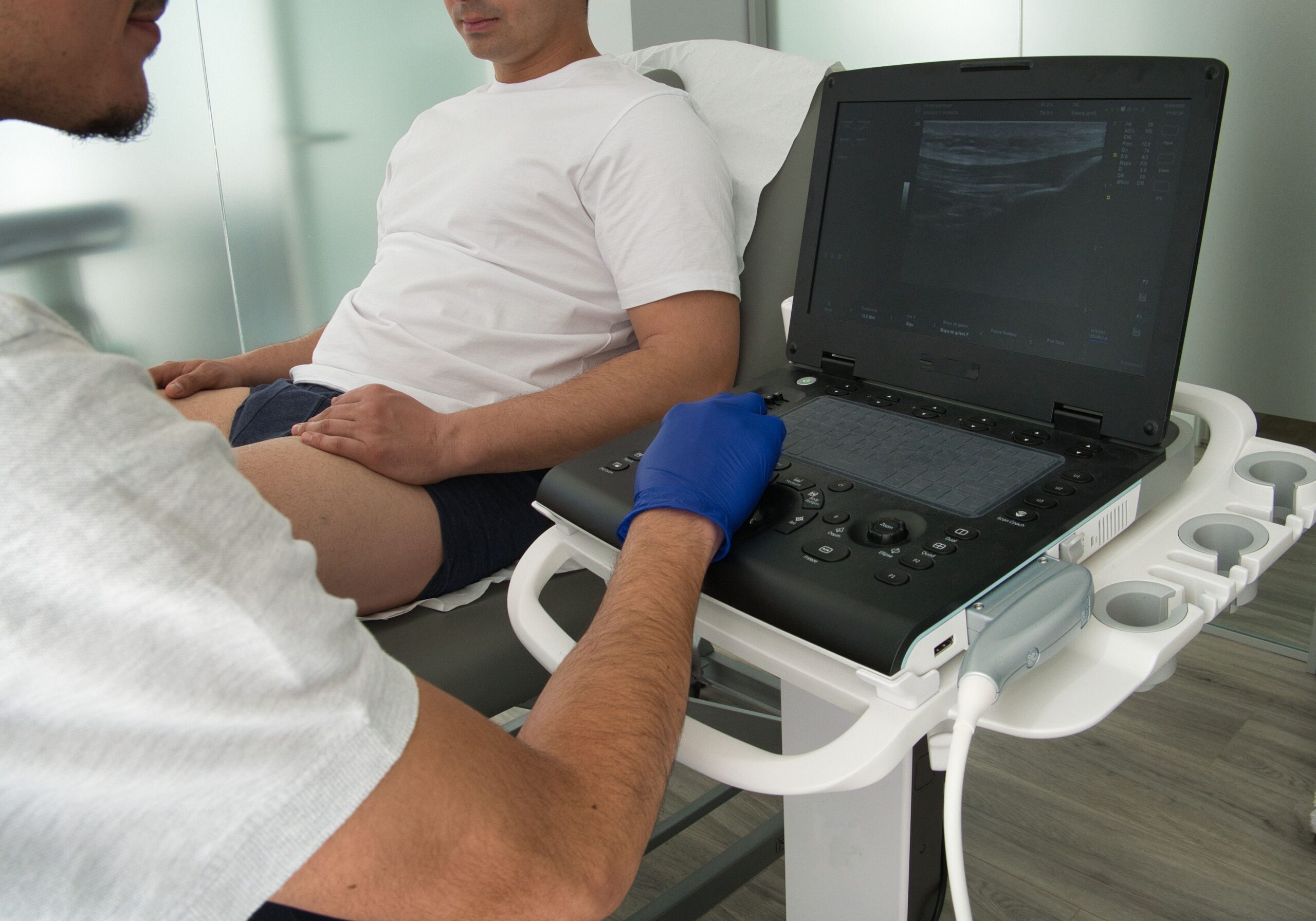
The procedure for a prostate gland ultrasound is performed with attention to patient comfort and imaging accuracy. Here’s a step-by-step breakdown of what typically occurs during the ultrasound:
Preparation and Positioning
- Patient Positioning: The patient is usually positioned on the examination table, lying on his side with knees bent toward the chest to allow optimal access to the prostate gland.
- Preparation of Equipment: The ultrasound probe, or transducer, is covered with a protective sheath and lubricated adequately to ensure comfort during insertion.
Conducting the Ultrasound
- Insertion of the Transducer: The lubricated transducer is gently inserted into the rectum.
- Image Acquisition: The urologist moves the transducer to various positions to capture images of the prostate and the surrounding tissues. This process is typically painless, though it may cause some discomfort or a sensation of pressure.
- Real-Time Observation: Throughout the procedure, images are displayed on a monitor, allowing the practitioner to examine the prostate gland from different angles and check for abnormalities.
- Image Saving: Some captured images are saved digitally for detailed post-procedure analysis and for maintaining a record that can be referenced in future follow-ups.
Duration
The entire procedure usually takes about 20 to 30 minutes, depending on the complexity of the case and the quality of the images obtained.
Interpreting Results
Interpreting the results of a prostate gland ultrasound involves a detailed analysis of the images obtained during the procedure. Here’s how the results are generally interpreted:
Understanding Normal Findings
- Prostate Size and Shape: The normal prostate gland has a symmetrical shape and size, typically measuring about 20 to 30 grams in volume.
- Echo Texture: A homogeneous echotexture is usually indicative of a healthy prostate.
Identifying Abnormal Findings
- Lesions or Masses: Hypoechoic (darker) areas within the prostate may be indicative of prostate cancer, especially if located in the peripheral zone. Hyperechoic (brighter) areas might represent calcifications or chronic inflammation.
- Vascularity: Increased blood flow in areas within the prostate can be a sign of malignancy. Normal prostate tissue typically shows minimal vascularity.
Implications of Findings
- Benign Conditions: Enlarged prostate or benign lesions usually require monitoring and potential medical management depending on the severity of symptoms.
- Suspicious Findings: Areas that appear suspicious for malignancy often necessitate further investigation, typically through a guided biopsy to confirm the presence of cancer cells.
- Inflammatory Conditions: Signs of inflammation may lead to additional testing or treatment for prostatitis.
Risks and Considerations
While prostate gland ultrasound is a generally safe and non-invasive procedure, there are some potential risks and considerations:
- Minor Bleeding: Inserting the ultrasound probe into the rectum can cause minor irritation or damage to the rectal lining, resulting in slight bleeding. If an ultrasound-guided biopsy is performed, there is also a small risk of bleeding from the biopsy site within the prostate.
- Discomfort and Pain: The insertion and manipulation of the ultrasound probe in the rectum can cause a sense of pressure or discomfort.
- Risk of Infection: Any procedure involving inserting an instrument into the body risks introducing pathogens. Although rare, there is a slight possibility of infection following a prostate ultrasound, particularly if a biopsy is taken.
Conclusion
Prostate gland ultrasound is a diagnostic tool in urology that offers a non-invasive method to assess the prostate and guide further diagnostic or therapeutic actions.
While generally safe, patients need to be aware of the minor risks associated with the procedure, such as discomfort, slight bleeding, and rare instances of infection. These considerations should be balanced against the significant benefits of accurate diagnosis and guided treatment.
For those seeking further information or wishing to schedule a prostate ultrasound, visit our clinic’s website to find out more.