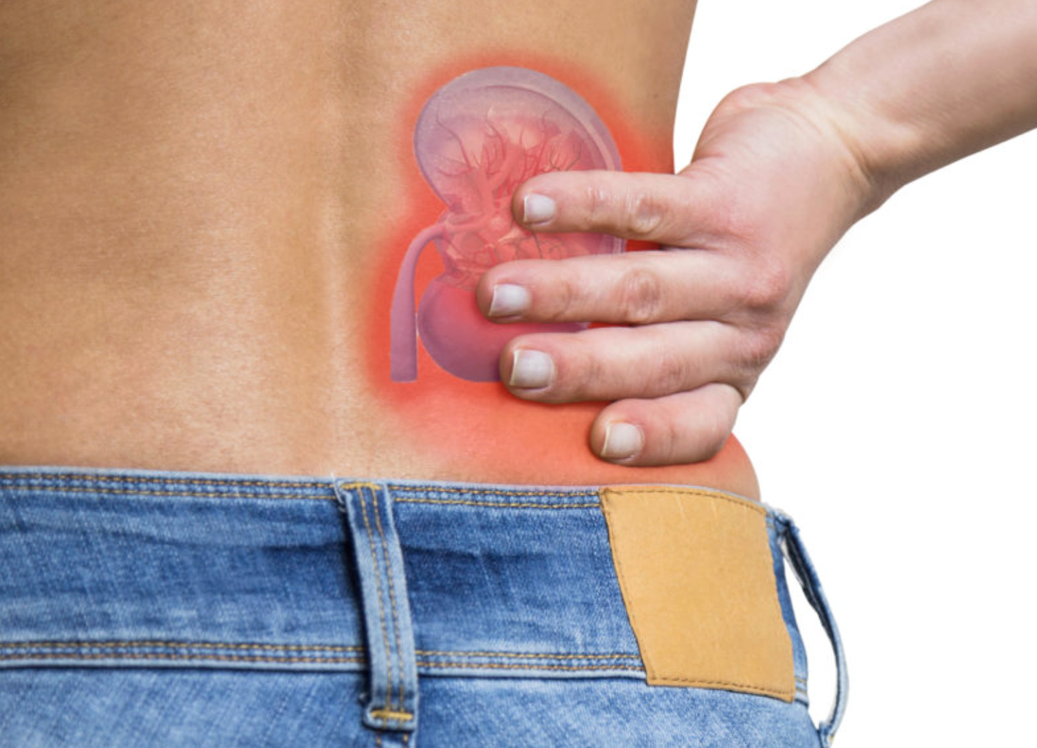
Can you tell the difference between a muscle strain and a kidney stone emergency? Renal colic pain begins without warning, often in the early morning hours as a dull flank ache that rapidly escalates to severe, cramping pain radiating toward the groin. The pain occurs when a kidney stone moves from the kidney into the ureter (the tube that carries urine from the kidney to the bladder). The muscular tube spasms as it attempts to push the stone toward the bladder. Unlike other types of abdominal pain, where staying still provides relief, renal colic sufferers often find themselves unable to find any comfortable position. Pacing, rocking, or constantly shifting provides the only marginal relief from the waves of intense discomfort.
The pain pattern follows the stone’s journey through the urinary tract. Upper ureteral stones produce flank and upper abdominal pain. Mid-ureteral stones cause pain radiating to the lower abdomen. Stones near the bladder junction trigger urinary urgency, frequency, and pain radiating to the groin or genitals. Recognising these patterns helps determine the stone’s location and guides treatment decisions.
Recognising Renal Colic Symptoms
Renal colic presents with distinctive characteristics that differentiate it from other abdominal emergencies. The pain arrives in waves, with varying durations and periods of reduced intensity between episodes. Nausea and vomiting commonly accompany severe pain due to shared nerve pathways between the kidney and gastrointestinal tract.
Visible blood in urine or tea-coloured urine occurs when the stone’s rough edges irritate the ureteral lining. Some patients notice only microscopic blood detected on urine testing. Urinary symptoms intensify as the stone approaches the bladder. Burning during urination, a persistent urge to urinate despite passing small amounts, and lower abdominal pressure become prominent.
Fever and chills signal a potential urinary tract infection complicating the stone obstruction. This combination represents an emergency requiring immediate medical attention. Infected urine trapped behind a stone can rapidly progress to sepsis (a life-threatening response to infection).
Pain Location Patterns
Stones lodged in the upper ureter cause flank pain, often mistaken for muscle strain. The pain may radiate around the side toward the front of the abdomen. Mid-ureteral stones produce pain that follows the ureter’s course. The pain tracks from the flank toward the lower abdomen on the affected side.
Lower ureteral stones near the bladder junction create symptoms mimicking urinary tract infections. These include urgency, frequency, and suprapubic discomfort (pressure above the pubic bone). Men may experience testicular pain, while women may feel discomfort radiating to the labia. These referred pain patterns occur because the lower ureter shares nerve supply with pelvic structures.
Immediate Actions When Pain Begins
Heat application to the flank and lower back can provide pain relief. It helps relax the ureteral smooth muscle and reduce the intensity of spasm. A heating pad set to medium heat or a hot water bottle wrapped in a towel, applied for appropriate intervals, offers non-pharmacological pain control while awaiting medical care or medication onset.
Hydration during acute renal colic remains a topic of discussion among urologists (doctors who specialise in urinary tract conditions). Adequate fluid intake helps move stones through the urinary tract. However, forcing excessive amounts of fluid during acute obstruction may worsen pain by increasing pressure above the stone. Moderate fluid intake represents a balanced approach. Sip water regularly rather than drinking large volumes rapidly.
Movement and position changes help many patients cope with pain waves. Walking, gentle stretching, or rocking in a chair may provide psychological distraction and physical comfort. Some patients find relief sitting forward in a warm shower with water directed at the affected flank.
Over-the-Counter Pain Management
Non-steroidal anti-inflammatory drugs (NSAIDs) like ibuprofen can provide renal colic relief. They help reduce inflammation and decrease ureteral spasm. NSAIDs work through a different mechanism than pure analgesics. They directly address the underlying ureteral inflammation that generates pain signals. Take with food to minimise gastric irritation.
Paracetamol offers modest pain relief. You can combine it with NSAIDs for an additive effect when single agents provide insufficient control. The combination allows lower doses of each medication while achieving adequate pain management.
Antispasmodic medications (medicines that reduce muscle cramping) available over the counter may help reduce the cramping quality of renal colic. Evidence for their effectiveness specifically in kidney stone pain remains limited.
When to Seek Professional Help
- Fever above 38°C accompanying flank pain
- Inability to keep fluids down due to persistent vomiting
- Pain uncontrolled by maximum doses of over-the-counter medications
- Complete inability to pass urine
- Known single kidney or previous kidney surgery
- Pregnancy with suspected renal colic
- Pain lasting more than several hours without improvement
💡 Did You Know?
The ureter’s diameter measures only a few millimetres at its narrowest points. These include the ureteropelvic junction (where the ureter meets the kidney), where it crosses the iliac vessels, and the ureterovesical junction entering the bladder. Smaller stones pass spontaneously in many cases, while larger stones rarely pass without intervention.
What Happens During Emergency Evaluation
Emergency assessment begins with vital signs (such as blood pressure, heart rate, and temperature), urine testing, and blood tests checking kidney function and infection markers. Urine analysis reveals blood cells in many cases of renal colic. It may show white blood cells, indicating infection, or crystals, suggesting stone composition.
A non-contrast CT scan (a specialised imaging test that produces detailed images of the urinary tract) is the definitive imaging study. It can identify very small stones and reveal their precise location, size, and any associated complications, such as hydronephrosis (kidney swelling from urine backup). CT also excludes other conditions mimicking renal colic, including appendicitis, ovarian pathology, and abdominal aortic aneurysm.
Pain control in the emergency setting typically involves intravenous ketorolac (an NSAID) or opioid medications for severe, refractory pain. Intravenous fluids address dehydration from vomiting and reduced oral intake. Antiemetic medications (medicines that prevent nausea and vomiting) control nausea. This improves comfort and facilitates the transition to oral medication.
Decisions About Admission vs. Discharge
Many patients with uncomplicated renal colic return home with pain medications, anti-nausea medications, and a strainer to catch passed stones for analysis. Outpatient urology follow-up occurs within a short time frame if the stone hasn’t passed.
Hospital admission becomes necessary in several situations:
- When infection complicates obstruction
- When kidney function deteriorates
- When intractable vomiting prevents oral intake
- When adequate pain control proves difficult with oral medications
Patients with stones in a solitary kidney require closer monitoring due to the risk of complete renal obstruction.
Medical Expulsive Therapy
Alpha-blocker medications like tamsulosin relax smooth muscle in the lower ureter. They can increase stone passage and shorten passage time. Initially developed for prostate conditions, these medications dilate (widen) the narrow ureterovesical junction where stones commonly lodge.
A healthcare professional may prescribe tamsulosin for distal ureteral stones (stones in the lower part of the ureter near the bladder) under a specific size where observation seems reasonable. The medication is typically taken once daily, usually at bedtime, to minimise dizziness from blood pressure effects. Treatment continues until stone passage or scheduled intervention.
Adequate analgesia (pain relief) during the waiting period helps prevent suffering and reduce emergency department returns. A combination prescription of scheduled NSAIDs with breakthrough opioid medication for severe pain episodes represents typical outpatient management.
⚠️ Important Note
Severe renal colic pain should be managed with appropriate medication. Uncontrolled pain triggers physiological stress responses. These responses can elevate blood pressure, worsen nausea, and delay stone passage by increasing ureteral spasm.
Interventions for Stones That Won’t Pass
Stones failing to progress after several weeks of medical management may require urological intervention. Stones too large for spontaneous passage may also require intervention. A healthcare professional will select the procedure based on:
- Stone size
- Stone location
- Stone composition (if known)
- Patient anatomy
Shock wave lithotripsy uses focused acoustic waves to fragment stones into passable pieces without incisions. Healthcare providers perform it as an outpatient procedure. Patients typically return home the same day and pass fragments over the following weeks. Stones in the upper ureter and kidney respond to this technique.
Ureteroscopy involves passing a thin scope through the urethra and bladder into the ureter. The doctor visualises the stone directly and fragments it with laser energy. The doctor removes stone fragments during the procedure. A temporary stent may remain to ensure urine drainage while ureteral swelling resolves.
Percutaneous nephrolithotomy addresses large kidney stones through a small flank incision. This procedure accesses the kidney directly to remove a substantial stone burden. This procedure requires brief hospitalisation but offers complete stone clearance for complex cases.
Prevention After the Acute Episode
Stone composition analysis guides targeted prevention strategies. Calcium oxalate stones, a common type, respond to dietary modifications. These include:
- Moderate calcium intake from food sources
- Reduced sodium consumption
- Limited oxalate-rich foods like spinach, rhubarb, and nuts
Uric acid stones form in acidic urine. They may dissolve with urinary alkalinisation using potassium citrate supplements. Dietary purine reduction helps decrease uric acid production. This means limiting red meat, organ meats, and certain seafood.
Adequate hydration remains a prevention measure across all stone types. Target urine output to achieve a pale yellow colour throughout the day. This helps dilute stone-forming substances and reduce the risk of crystallisation.
✅ Quick Tip
Save any passed stone or stone fragments by urinating through a fine strainer. Rinse the stone. Place it in a clean container and bring it to your urology appointment. Stone composition analysis allows personalised prevention recommendations rather than generic dietary advice.
Managing Recurrent Episodes
Patients with previous kidney stones face an elevated recurrence risk, particularly without preventive measures. Metabolic evaluation using appropriate urine collection can identify specific abnormalities that drive stone formation. These include excess calcium excretion, low citrate levels, high oxalate, or abnormal urine pH.
Based on metabolic findings, a healthcare professional may recommend dietary modifications alone or add specific medications:
- Thiazide diuretics reduce calcium excretion in recurrent calcium stone formers
- Potassium citrate increases urinary citrate (a natural stone inhibitor)
- Potassium citrate alkalinises urine for uric acid stone prevention
Regular imaging surveillance monitors for new stone formation before symptoms develop. Healthcare providers may electively treat small stones identified early, when convenient, rather than emergently when they cause acute obstruction.
Commonly Asked Questions
How long does a kidney stone take to pass?
Response times vary depending on individual circumstances. Smaller stones typically pass more quickly with adequate hydration and medical expulsive therapy. Moderately sized stones may take longer. Larger stones often require intervention, though passage remains possible. Location matters significantly. Stones already in the lower ureter pass faster than those in the upper ureter or kidney.
Can I prevent another kidney stone episode?
Recurrence risk can decrease substantially with appropriate prevention. Hydration targeting clear to pale yellow urine throughout the day can reduce recurrence. Dietary modifications based on stone composition and metabolic evaluation can further reduce risk. Some patients may benefit from preventive medications when dietary measures prove insufficient.
Is it safe to wait for a stone to pass instead of having surgery?
Observation remains appropriate for uncomplicated stones of specific sizes when pain is manageable and no infection is present. However, prolonged obstruction beyond several weeks risks permanent kidney damage from back-pressure. Regular follow-up imaging ensures the stone progresses and kidney swelling resolves rather than worsens.
What causes kidney stones to form?
Stone formation results from supersaturation of urine with crystal-forming substances. These include calcium, oxalate, uric acid, or other minerals. Several factors contribute:
- Inadequate fluid intake
- Dietary factors
- Genetic predisposition
- Certain medications
- Metabolic conditions
Identifying individual risk factors through testing allows targeted prevention.
Will drinking cranberry juice help pass my kidney stone?
Cranberry juice does not help pass existing stones. It may actually worsen calcium oxalate stone formation by increasing urinary oxalate. While cranberry products may help prevent urinary tract infections, they offer no benefit for the management of kidney stones. Plain water remains the preferred fluid for stone patients.
Next Steps
Apply heat to the flank for immediate pain relief and use NSAIDs as first-line pain management. Seek emergency care if you develop a fever above 38°C, persistent vomiting, or complete inability to urinate. Once the acute episode resolves, obtain stone composition analysis to guide personalised prevention strategies, including adequate hydration and targeted dietary modifications.
If you’re experiencing severe flank pain radiating to the groin, blood in your urine, or recurrent kidney stone episodes, schedule an evaluation with a urologist for diagnostic imaging and personalised treatment planning.