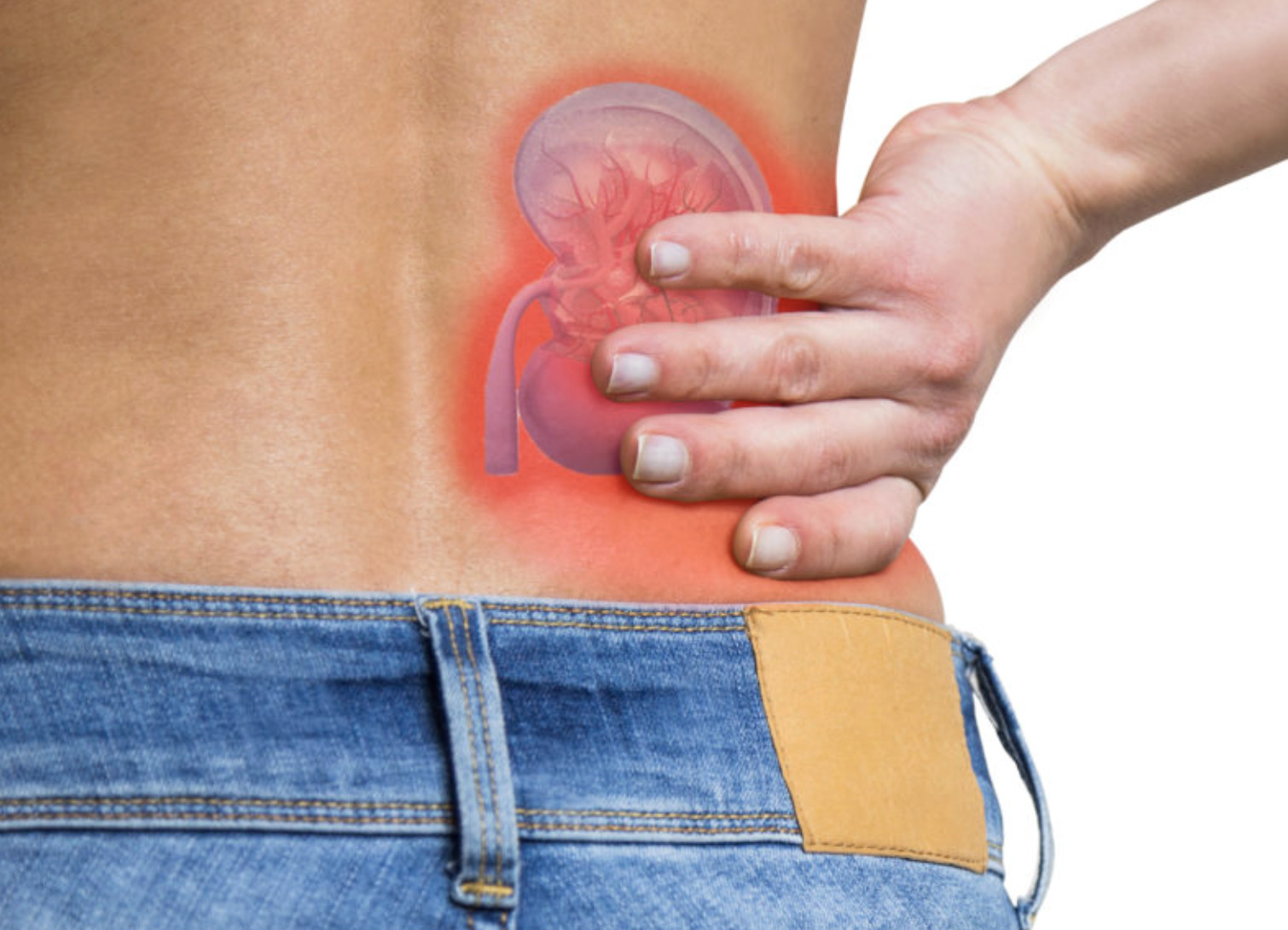
Can a kidney stone exist in your body for years without causing a single symptom? Kidney stones discovered incidentally during imaging for unrelated conditions present a clinical dilemma. These silent stones—causing no pain, bleeding, or urinary symptoms—exist in many adults who remain entirely unaware of them until a CT scan or ultrasound reveals their presence. The question of whether to treat or monitor these asymptomatic stones depends on multiple factors. These include size, location, composition, and individual patient characteristics.
Not all kidney stones behave the same way. Some remain dormant for years, never causing symptoms or complications. Others grow silently until they obstruct urine flow or trigger sudden, severe pain.
How Silent Kidney Stones Are Discovered
Most asymptomatic stones appear on imaging performed for other medical evaluations. Abdominal CT scans (a type of imaging test that uses X-rays to create detailed cross-sectional images of the body) for appendicitis, trauma assessment, or cancer screening frequently reveal incidental renal calculi. Ultrasound examinations (a test that uses sound waves to create images of internal organs) for liver, gallbladder, or gynaecological concerns may detect stones as echogenic foci with acoustic shadowing.
Preoperative imaging before abdominal or pelvic surgery can detect previously unknown stones. Health screening programmes, including comprehensive body scans, identify stones in individuals with no urinary complaints whatsoever.
Once discovered, these stones require evaluation even without symptoms. Stone characteristics visible on imaging—size, location, density, and number—provide initial information for management planning.
Factors Determining Treatment Necessity
Stone Size and Growth Potential
Size remains a primary factor in treatment decisions for silent stones. Stones with a diameter less than a specific threshold rarely cause obstruction. They often pass spontaneously if they do enter the ureter (the tube that carries urine from the kidney to the bladder). These small stones may warrant observation alone, with periodic imaging to monitor for growth.
Stones in an intermediate size range occupy an area where individual factors influence decisions. Location within the kidney matters. Stones in the lower pole may grow without causing symptoms. Those near the ureteropelvic junction (where the kidney connects to the ureter) pose a higher risk despite smaller size.
Larger stones rarely pass spontaneously. They carry an increased risk of complications, including infection and progressive kidney damage. Larger stones also tend to grow faster than smaller ones.
Stone Location and Anatomy
Where a stone sits within the collecting system (the kidney’s drainage network) affects both the risk of symptoms and the treatment approach. Upper and middle pole stones may pass into the ureter more readily than lower pole stones. Lower pole stones can grow substantially while remaining trapped in dependent calyces (cup-shaped structures within the kidney that collect urine).
Kidney anatomy varies between individuals. Some people have narrow infundibula—the passages connecting calyces to the central renal pelvis—that prevent even moderate-sized stones from migrating. Others have capacious collecting systems where stones move freely.
Stones in solitary kidneys (when a person has only one functioning kidney) or in transplanted kidneys require more careful treatment. Any obstruction in a single functioning kidney threatens total renal function.
Stone Composition Clues
CT scan density, measured in Hounsfield units (a scale that indicates how dense a material appears on a CT scan), provides clues about stone composition. Lower density stones often contain uric acid (a waste product that can crystallise in urine). They may respond to urinary alkalinisation (raising urine pH to reduce acidity) without surgical intervention. Higher-density stones typically contain calcium. They require mechanical removal if treatment becomes necessary.
Pure uric acid stones can sometimes dissolve completely with oral medication that raises urine pH above a certain threshold. Dual-energy CT scanning can differentiate stone types with reasonable accuracy when available.
Calcium oxalate and calcium phosphate stones—the most common types—do not dissolve with medical therapy. However, metabolic evaluation (testing to identify factors that promote stone formation) and dietary modification can slow their growth.
Risks of Observation Versus Intervention
What Happens Without Treatment
Natural history studies following patients with untreated asymptomatic stones show variable outcomes. Some stones remain stable for years without causing problems. Others grow progressively or eventually cause acute symptoms requiring emergency intervention.
Silent stones can cause subtle damage not apparent without specific testing. Chronic partial obstruction may gradually impair kidney function. Stone-related infection can develop without warning, potentially progressing to serious systemic illness.
Larger stones occasionally cause painless haematuria—blood in urine—that patients may not notice without urinalysis (a laboratory test that examines urine for various substances and abnormalities). Persistent microscopic bleeding warrants investigation.
💡 Did You Know?
Kidney stones can serve as a nidus (a central point where bacteria gather) for bacterial biofilm formation. This harbours infections that resist antibiotic treatment. Removing the stone eliminates this bacterial reservoir completely.
Procedural Considerations
Modern stone treatment carries low but non-zero risks. Shock wave lithotripsy—breaking stones with focused sound waves—requires no incisions. However, it may cause temporary bleeding or incomplete fragmentation. Ureteroscopy (a procedure in which a doctor inserts a thin, flexible tube with a camera into the urinary tract to visualise and treat stones) provides direct visualisation and laser fragmentation. It requires anaesthesia and carries small risks of ureteral injury.
Percutaneous nephrolithotomy (a procedure where the doctor creates a small passage through the skin directly into the kidney to remove stones) treats larger stones. This approach offers high clearance rates but involves greater procedural complexity and recovery time. Choosing between observation and any intervention requires weighing these procedural risks against the risks of leaving stones untreated.
Patient factors influence procedural risk substantially. Anticoagulation therapy (blood-thinning medication), bleeding disorders, active infection, and anaesthesia concerns all affect treatment safety.
Surveillance Protocols for Monitored Stones
When you choose observation, structured follow-up ensures any concerning changes are timely reassessed. Imaging intervals depend on initial stone characteristics and individual risk factors.
Small stones in low-risk patients typically warrant initial imaging at regular intervals. Stable stones on two or more imaging studies may warrant less frequent monitoring. Any growth, new symptoms, or concerning laboratory findings accelerate the timing of follow-up.
Metabolic evaluation (comprehensive testing to identify the specific factors that cause stones to form in your body) identifies correctable factors that promote stone formation. Twenty-four-hour urine collections (where you collect all urine produced over a full day for analysis) measure calcium, oxalate, citrate, uric acid, and other relevant parameters. Dietary and medical interventions based on these results can slow stone growth during observation periods.
⚠️ Important Note
Patients with observed kidney stones should seek prompt evaluation for new flank pain, visible blood in urine, fever, or difficulty urinating. These symptoms may indicate stone movement or complication requiring urgent assessment.
Medical Management During Observation
Dietary Modifications
Fluid intake represents one of the more impactful dietary changes for stone prevention. Maintaining substantial urine output daily dilutes stone-forming substances. It promotes the passage of small crystals before they aggregate. Water remains the preferred fluid. Citrus juices provide additional citrate (a natural substance that inhibits stone formation) that helps prevent stones from developing.
Sodium restriction reduces urinary calcium excretion (the amount of calcium released in urine) regardless of dietary calcium intake. Processed foods, restaurant meals, and added table salt contribute substantially to sodium load. Moderating sodium intake supports stone prevention without requiring extreme dietary limitations.
You should get dietary calcium from food rather than supplements. Adequate calcium binds oxalate (a compound found in many foods that can contribute to stone formation) in the intestine. This prevents its absorption and subsequent excretion in urine. Low-calcium diets paradoxically increase the risk of kidney stones by allowing greater oxalate absorption.
Medications That Slow Stone Growth
Thiazide diuretics (medications that help the kidneys remove excess fluid whilst reducing urinary calcium excretion) reduce urinary calcium excretion. They can benefit patients with documented hypercalciuria (excessively high urinary calcium levels) on metabolic testing. These medications require monitoring for electrolyte changes, but can slow calcium stone growth when indicated.
Potassium citrate increases urinary citrate—a natural stone inhibitor—and raises urine pH (makes urine less acidic). This dual action can benefit multiple stone types. The alkalinising effect can dissolve existing uric acid stones whilst helping prevent new stone formation.
Allopurinol (a medication that reduces the body’s production of uric acid) reduces uric acid production. It can benefit patients with hyperuricosuria (elevated uric acid levels in urine), whether forming uric acid or calcium oxalate stones. Elevated urinary uric acid promotes calcium oxalate crystallisation through complex chemical interactions.
When Silent Stones Mandate Treatment
Absolute Indications
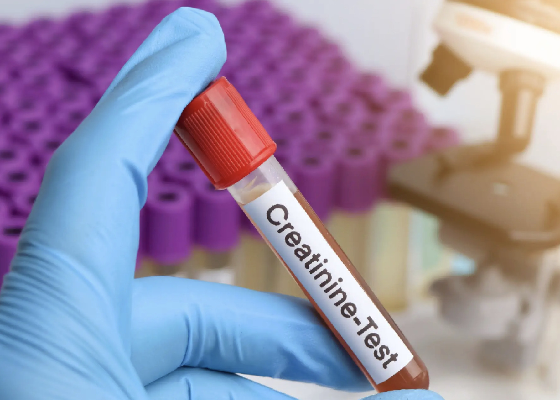
Certain situations require treatment regardless of symptom status. Stones causing progressive decline in kidney function—documented by comparing creatinine levels (a waste product that indicates how well the kidneys are filtering) or nuclear medicine scans over time—warrant intervention to preserve renal parenchyma (the functional kidney tissue).
Stones associated with chronic urinary tract infections, particularly those caused by urea-splitting organisms (bacteria that produce enzymes breaking down urea into ammonia), require complete removal. These infection stones, often called struvite stones, grow rapidly. They can fill the entire collecting system if untreated.
Stones in patients requiring immunosuppression (medications that reduce immune system activity), chemotherapy, or other treatments that impair the infection-fighting ability should generally be removed. The risk of complicated infection during immune compromise outweighs the procedural risks of stone treatment.
Occupational and Lifestyle Considerations
Certain occupations make unpredictable stone symptoms particularly problematic. Pilots, military personnel, long-haul truck drivers, and others in safety-sensitive positions may benefit from elective stone treatment to prevent symptoms during activities.
Patients planning extended travel to areas with limited medical facilities face similar considerations. Developing ureteral colic (sudden, severe pain caused by a stone blocking the ureter) in a remote location creates significant hardship and potential danger. Elective treatment before such travel eliminates this risk.
Women planning pregnancy should discuss stone management beforehand. Symptomatic stones during pregnancy limit treatment options and complicate care. Addressing known stones before conception simplifies management considerably.
What Our Urologist Says
Stone management decisions balance multiple factors unique to each patient. A small stone in a healthy young person with no metabolic abnormalities often warrants observation with lifestyle modifications. The same stone in someone with recurrent infections, anatomical abnormalities, or occupational constraints might be more appropriate for intervention. Your doctor can provide personalised recommendations based on your specific risk factors, overall health status, and individual circumstances.
Modern endoscopic techniques (procedures using thin tubes with cameras to examine and treat internal structures) have reduced treatment morbidity substantially compared to historical approaches. For appropriately selected patients, recovery from stone treatment is brief. Complications are uncommon.
Shared decision-making remains essential. Patients who understand their stone characteristics, natural history expectations, and treatment options can participate meaningfully in choosing between observation and intervention. Neither approach suits every situation—individual circumstances guide appropriate recommendations.
Putting This Into Practice
- Maintain daily fluid intake sufficient to produce pale or clear urine throughout the day.
- Reduce sodium in your diet by limiting processed foods and avoiding added salt.
- Continue eating calcium-rich foods such as dairy products, leafy greens, and fortified alternatives.
- Complete any metabolic testing your urologist recommends to identify specific stone risk factors.
- Keep scheduled follow-up imaging appointments even when feeling completely well.
When to Seek Professional Help
- New flank pain or discomfort in the side or back
- Visible blood in urine, or urine appearing pink, red, or brown
- Fever or chills, which may indicate stone-related infection
- Persistent nausea or vomiting
- Difficulty urinating or significantly reduced urine output
- Burning or pain during urination
Commonly Asked Questions
Can kidney stones dissolve on their own without treatment?
Uric acid stones can dissolve with urinary alkalinisation using potassium citrate, typically over several months. Calcium-containing stones—the most common type—do not dissolve with any medication. They require mechanical removal if treatment becomes necessary.
How quickly do kidney stones typically grow?
Growth rates vary considerably based on metabolic factors and stone composition. Some stones remain stable for years. Others enlarge substantially within months. Serial imaging identifies individual growth patterns and informs management decisions.
Will a silent kidney stone eventually cause pain?
Not necessarily. Some asymptomatic stones remain in the kidney indefinitely. Others eventually migrate towards the ureter, causing pain when they obstruct urine flow. Predicting which stones will become symptomatic remains imprecise.
Is it safe to exercise with a known kidney stone?
Regular physical activity is generally safe. It may even help prevent additional stone formation. However, activities that pose a significant risk of dehydration—prolonged endurance exercise, hot yoga, sauna use—require attention to fluid replacement.
How often should I have imaging to monitor my kidney stone?
Initial monitoring may be recommended at regular intervals. Stable stones across multiple studies may transition to annual or less frequent surveillance. Your healthcare provider can establish monitoring schedules tailored to your individual risk factors based on your stone characteristics. Any new symptoms warrant immediate reassessment regardless of scheduled follow-up timing.
Next Steps
Silent kidney stones require individualised evaluation based on size, location, and patient-specific factors. Regular surveillance with metabolic optimisation supports safe monitoring when appropriate. Stones causing progressive kidney damage, recurrent infections, or occurring in high-risk situations warrant intervention.
If you’ve been told you have a kidney stone on imaging, or if you’re experiencing flank pain, blood in urine, or recurrent urinary infections, consult a urologist for a comprehensive assessment and personalised management recommendations.